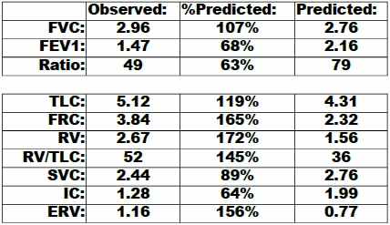
VA, DLCO and COPD

Although the technology used to perform the single-breath DLCO test has improved since it was first developed in the 1950’s the essential concepts and equations have not changed significantly. Probably the most important advance has been the introduction of rapid response real-time gas analyzers in the 1990’s. Prior to that time the patient’s washout and sample volumes had to be preset which always involved a certain amount of guesswork when a patient was significantly obstructed or restricted. With a real-time gas analyzer it is possible inspect the exhaled gas tracings after the test has been performed in order to determine when washout has occurred and then select the appropriate location for the sample volume. This has improved the single-breath DLCO test quality but at the same time it has also exposed some of its limitations.

The single-breath DLCO test attempts to simplify what is actually a very complex process. One of the key assumptions of the single-breath DLCO calculations is that the inspired gas mixture is evenly distributed throughout the lung. This is not really true even for patients with normal lungs and in general, inspired gas follows the last in-first out rule. In patients with lung disease this inhomogeneous filling and emptying can be magnified and a maldistribution of ventilation is often most evident in patients with COPD.

Continue reading

Busted for speeding during an N2 washout

Nitrogen washout lung volumes are still relatively new to my PFT Lab. The number of problems we’ve encountered has decreased substantially but we are still learning some of the idiosyncrasies of the system. Recently while trying to understand a test with odd results we were reminded by the manufacturer that during the washout period a patient’s inspiratory and expiratory flow rates should not exceed 1.5 liters/second. The reason this “speed limit” is necessary highlights some of the limitations of modern open-circuit lung volume measurements.

The basic concept behind nitrogen washout is relatively simple. The air we breathe contains 78% nitrogen which is a relatively inert, insoluble gas. If you have a patient breathe 100% oxygen and then collect their exhaled air you can calculate the volume of exhaled nitrogen by multiplying the concentration in the exhaled air by the total volume of air that was collected. Once you know the volume of nitrogen you can then calculate the lung volume.

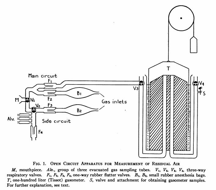
Initially this was a laborious and cumbersome process. The patient’s exhaled breathing circuit and a Tissot Gasometer (a very large spirometer with a volume between 125 and 300 liters) are first flushed with oxygen several times to remove any nitrogen. Next, while breathing room air the patient exhales to RV and an end-expiratory gas sample is taken and used to estimate the patient’s alveolar nitrogen concentration. The patient is then switched to 100% oxygen and breathes for seven minutes. At the end of the washout period the nitrogen concentration of the exhaled gas in the gasometer is analyzed and the volume recorded.

 N2_Washout_System_JCI_v19_n4_p609_1940

Continue reading

N2 washout switch-in error

I have been taking a close look at the raw data from all lung volume tests lately in large part because N2 washouts are still relatively new to my PFT Lab and we’re continuing to learn from our mistakes. When I saw this N2 washout test I knew that there was something wrong with it. The patient had performed three N2 washout tests and the TLC, FRC and RV for this one test were significantly larger than for the other tests. The most common problem we’ve been having with N2 washouts has been with patient leaks during the washout period which almost always show up as an upwards drift in the tidal baseline. This test did not show any drift however, and it took me a little while before I could see what was wrong with it.

The N2 washout maneuver has the patient start by breathing tidally for a short period of time in order to determine where end-exhalation (FRC) is located. The patient then performs a slow vital capacity maneuver by steadily inhaling maximally to TLC and then exhaling maximally to RV. The technician then switches the patient into the washout breathing circuit at maximal exhalation and the patient resumes breathing tidally for the remainder of the test.

Continue reading

Lung volume measurements and software smarts.

Many years ago I used to think that lung volume measurements were the easy part of PFTs. As time has gone on I’ve seen that getting accurate lung volume measurements is actually more difficult than getting accurate spirometry and DLCO results mostly because the errors tend to be subtle.

The errors that occur in lung volume measurements tend to cause an overestimation of lung volumes. This often means that restrictive diseases can be unrecognized or that hyperinflation and gas trapping can be diagnosed where it does not exist.

I see questionable lung volume test results more often than I’d like even from experienced technicians. When I find what went wrong I try to use these as “teachable moments” for all the the lab staff. Despite this the number of questionable test results never seems to drop below a certain level. I’d much prefer the error level was zero but since this is a situation that involves humans making measurements on humans I am likely being overly optimistic. A more realistic goal is to ask that the testing systems be smarter.

Continue reading

Single-breath TLC measurements

I was reviewing the specifications of different pulmonary function test systems recently and saw that several manufacturers were advertising that some of their test systems are able to measure TLC, FRC and RV from a single-breath maneuver. This is true, but only to a very limited degree and I think it is reasonable to ask how accurate and clinically useful these measurements are and whether it is legitimate to bill for the test.

The measurement is made by having a patient exhale to RV and then inhale a gas mixture containing a tracer gas (an insoluble gas like helium or methane) to TLC. When the patient exhales, the degree by which the tracer has been diluted is then used to calculate the patient’s TLC. The math is quite simple and as is expressed as:

TLC = (inspired volume x (Fitrace/Fetrace)) – machine deadspace

This is done routinely as part of the DLCO test and there it is referred to as alveolar volume (VA) and it should be noted that all manufacturers are using the VA from the DLCO test as a substitute for TLC and are not performing single-breath TLC as a separate measurement.

Continue reading