Airway obstruction and the FVC

Spirometry is the most commonly performed (and mis-performed) pulmonary function test around the world. The apparent simplicity of spirometry is misleading since there are numerous subtleties that have a significant effect on the results.

I suspect that when the FVC is thought about it is most often considered to be an index towards the total capacity of the lung. That’s certainly true in it’s own way, but the FVC is actually a critically important factor when determining airway obstruction. I’ve had a number of reports across my desk lately where the patient had a reasonably large change in FVC when compared to their last visit but little change in FEV1, and this has made a difference in how the results are interpreted. For example:

Visit 1: Observed: %Predicted: Predicted:
FVC: 4.27 87% 4.91
FEV1: 3.36 84% 3.99
FEV1/FVC: 79 96% 82
Visit 2: Observed: %Predicted: Predicted:
FVC: 4.67 95% 4.91
FEV1: 3.38 85% 3.99
FEV1/FVC: 72 88% 82

Although the change in FVC is not significant by my lab’s standards (+0.40 L, +9%) and the FEV1 has hardly changed at all, the FEV1/FVC ratio has gone from being within normal limits to being under the LLN and therefore showing mild airway obstruction. Continue reading

Short efforts, gas trapping and leaks

Outside the pulmonary lab there is this notion that spirometry is supposed to be so easy that anyone can do it. You just tell the patient to take a deep breath in and blow out fast and to keep blowing until they’re empty. What’s so hard about that?

Sheesh. GIGO. I keep finding ways in which the patient, their physiology and our equipment can conspire in ways to produce errors that even experienced technicians can miss. I’ve been paying a lot of attention to flow-volume loops lately and maybe it’s for this reason that I’ve seen a steady stream of spirometry tests that have something wrong with the FVC volume.

What I’ve been seeing are flow-volume loops where the end of exhalation is to the left of either the start of the FVC inhalation or of the tidal loop. Taken at face value this means that the patient did not exhale as much as they inhaled (and that the FVC is therefore underestimated) but there are several reasons why this happens and it takes a bit of detective work to figure out the cause and what to do about it.

The simplest reason is a short expiratory time. Flow-volume loops however, do not show time, only flow and volume. Sometimes when a patient stops exhaling abruptly it’s easy to see that the effort is short.

Abrupt_Termination_02_FVL

Other times it isn’t as clear:

Short_Exhalation_FVL

and you need to look at the volume-time curve as well.

Continue reading

An FVC is not an SVC

I’ve discussed the issue of inserting a predicted FVC into the predicted lung volumes several times now. At the risk of beating this issue to death I’d like to put to rest the notion that an FVC and an SVC are the same thing.

A Forced Vital Capacity (FVC) maneuver is designed to measure the maximum expiratory flow rates, in particular the expired volume in 1 second (FEV1). It has long been recognized that the effort involved in the FVC maneuver can cause early airway closure, even in individuals with normal lungs, and that for this reason the vital capacity can be underestimated due to gas trapping. This effect is usually magnified with increasing age and in individuals with obstructive lung disease.

A Slow Vital Capacity (SVC) maneuver is designed to measure the lung volume subdivisions Inspiratory Capacity (IC) and Expiratory Reserve Volume (ERV), and to maximize the measured volume of the vital capacity. Due to the more relaxed nature of the SVC maneuver there is significantly less airway closure and for this reason the SVC volume is usually larger than the FVC, again even in individuals with normal lungs.

Comparing individual reference equations can be difficult but in general the reference equations for SVC and FVC agree with this. Taking the available SVC and FVC reference equations (unfortunately limited to Caucasian because there are almost no SVC equations for other ethnicities) it is apparent that the average predicted SVC is larger than the average predicted FVC, and that the magnitude of this difference increases with age:

SVC_vs_FVC_Male

Continue reading

The ratio-nal approach to predicted TLC

I’ve been reading Miller et al’s Laboratory evaluation of Pulmonary Function which was published in 1987. That was an interesting time since PFT equipment manufacturers had mostly transitioned to computerized systems but there were still a lot of manual systems in the field. For this reason the book’s instructions are still oriented mostly around manual pulmonary function testing and there are numerous warnings about double-checking the results from automated systems.

The book includes extensive discussion on the calculations and formulas used for testing which makes it useful as a teaching resource. The authors were also very concerned about the correct way to run a PFT lab so there is a fair amount of discussion about staff requirements for education and training (including the medical director) and staff behavior and conduct. To this end each chapter includes extensive instructions on the proper way to perform tests and treat patients. Although the tone of this is somewhat dated and I’d like to say these kind of reminders shouldn’t be necessary, it doesn’t hurt to set a standard on the level of professionalism we should aspire to.

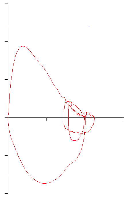
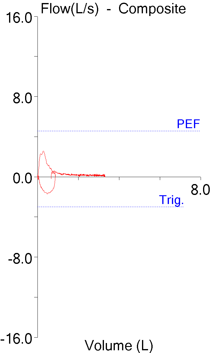
What caught my eye though, was a section in the chapter on Normal Values titled Interdependence of Normal Values which discussed of the value of deriving predicted TLC from predicted FVC. The authors were concerned that reference equations for different tests (and not just lung volumes) were being selected without concern for how well they fit together. I’ve previously written about the problems that results when inserting the reference equation for FVC into the reference equations for lung volumes. In one instance, the TLC was adjusted so that the final predicted TLC was equal to RV + VC, but this meant that TLC (and IC) were changed from the original reference equations. In another, the FVC was just substituted for SVC without adjustment which meant that RV + VC was not equal to TLC and IC + ERV was not equal to VC and this makes interpreting results problematic. What this means however, is that almost 30 years after this was published, this problem is still around.

As a solution, the authors point out that ratios, such as the FEV1/FVC ratio and the RV/TLC ratio tend to be relatively independent of height.

Since:

TLC = FVC + RV

This can be mathematically re-written as:

Which means that TLC can be derived from predicted FVC if the RV/TLC ratio is known.

Continue reading

The clue was in the O2

One of the overlooked parts of teaching pulmonary function interpretation is developing an appreciation for the number and variety of errors that the equipment, patients and technicians can produce and how they affect the reported test results. I routinely run across a couple dozen errors each week while reviewing reports. Most are minor and do not significantly affect the reported results. Many are mundane because they appear so often and a few are interesting because they point out a particular limitation in the equipment, software or testing standards. I’ve kept a file of the more iconic examples of testing errors for years and a while ago a pulmonary staff physician and I used to hold weekly sessions for fellows and residents where we’d present a number of “zingers” to see if they could figure them out. Unfortunately that physician has moved on to a different institution and I’m no longer as available as I used to be so these sessions are no longer held but I think that they or something like them should be held in all teaching hospitals.

These spirometry results came from a middle-aged woman with sarcoidosis.

Observed: %Predicted: Predicted:
FVC: 3.55 155% 2.29
FEV1: 1.06 60% 1.77
FEV1/FVC Ratio: 30 39% 77

Elevated FVC’s are not all that uncommon (and are a good example of the limitations of reference equations), but one that is 155% of predicted is particularly unusual. This occurs most commonly when somebody has made an error in measuring or entering the patient’s height (I can’t tell you the number of times I’ve seem someone entering 60 inches when they meant 6 feet), but this patient has been seeing pulmonary physicians and having regular spirometry tests for over a decade and the height for this test was the same as it was for the previous visit. In addition the trend report showed that over the last year the patient’s FVC had been between 71% and 65% of predicted.

Transtracheal_O2_FVL

The flow-volume loop doesn’t look overly unusual although the expiratory flow doesn’t taper off to zero and the patient maintained a low expiratory flow for at least two-thirds of the vital capacity.
Continue reading

Post-BD FVC. It’s about time.

When assessing the response to bronchodilators the change in FEV1 is used far more frequently than any other spirometry result. Other values such as inspiratory capacity (IC) and peak inspiratory flow (PIF) have also been proposed as indicators, but the ATS/ERS standards includes changes in FVC as well as changes in FEV1 and this is often overlooked. Specifically they:

…recommend using the per cent change from baseline and absolute changes in FEV1 and/or FVC in an individual subject to identify a positive bronchodilator response. Values >12% and 200 mL compared with baseline during a single testing session suggest a ‘‘significant’’ bronchodilatation.”

I don’t have any particular disagreement with this since post-BD increases in FVC are probably similar in nature to the post-BD changes in IC seen in some individuals with COPD. So when spirometry results like this:

Pre-BD: %Predicted: Post-BD: %Change:
FVC: 1.82 66% 2.55 +40%
FEV1: 0.66 32% 0.72 +10%
FEV1/FVC: 37 49% 29 -22%

comes across my desk, I’m inclined to consider that the results show a positive bronchodilator response. Post-BD increases in FVC are not usually quite as large as 40% however, so I took a closer look at this particular test. When I did what I saw was that the post-BD test length was significantly longer than the pre-BD test length.

Longer_FVC

Continue reading

Standing height, ethnicity and the vital capacity

In 1844 John Hutchinson published his first paper describing his spirometer and his research on the Vital Capacity. He was the first person to use the word “spirometer” to describe his instrument and the first to use the term “vital capacity” to designate the maximum amount of air an individual can exhale after a maximal inhalation. Although he is remembered as the inventor of the spirometer, he was not the first person to use a gasometer to measure lung volumes nor was he the first to measure the vital capacity. What made his research different from those that came before him was partly the prodigious number of individuals whose vital capacity he measured but far more importantly that he was able to show a clear relationship between standing height, age and vital capacity which had not been previously apparent. This finding galvanized researchers in England, Europe and the United States and in many ways helped set the course of research into lung function for many decades to come.

This clear relationship between standing height and vital capacity has been taken as scientific fact since that time despite inconsistencies not only in Hutchinson’s data but in almost all population studies since that time. The problem is that the relationship between standing height and vital capacity is not precise but only approximate. In order to explain the range of results that appeared in his data Hutchinson and other researchers of his time divided their study population into groups by their occupation. This approach may appear to be quaint to us now but at the time they were very serious both about the utility of doing this and what it told them about the different classes of society.

The first studies on vital capacity that divided the population by race were done in the United States. The reasons that this was done are both simple and complex, and overall there’s not a lot we can look back and be proud of. At that time there was an overwhelming societal concern with the races in general and not only the recently freed black slaves and the Amerindians but also about the different “races” of Europe that were emigrating to the United States. There was much public talk and private thought about the concepts of racial degeneracy, racial mongrelization and racial vitality, and unfortunately the vital capacity was taken as a way of measuring these things. Despite incredibly significant errors in both the methods and conclusions of these studies this approach spread to Europe during the second half of the 19th century and dividing study populations by race has become standard practice ever since.

When I first started doing pulmonary function testing I was taught to decrease the predicted vital capacity by 15% for Blacks and 10% for Asians. Decades later ethnicity-based population studies replaced these fractions. I always took this as the correct way to approach predicted values (and it is embedded in the ATS/ERS standards) but at the same time I’ve always had patients where it was either difficult to assign ethnicity or where their results significantly exceeded their ethnicity-based reference values. Over the last several years I have had the opportunity to study the issues surrounding reference equations extensively and I have become somewhat disenchanted with the notion of ethnicity-based reference equations.

Continue reading

When it’s FVC 1, EOT 2, volume comes out short

I was reviewing a pre- and post-bronchodilator spirometry report that showed a relatively large increase in FVC but the change in FEV1 was not significant. It’s not impossible for a patient to show this kind of a pattern following a bronchodilator but it is somewhat unusual. Usually when I see this it means that the patient exhaled a lot longer post-BD than they did pre-BD. When I looked however, I saw that just the opposite was true, the expiratory time was actually shorter for the post-BD effort than it was for the pre-BD effort.

FVC_Error_Table

The reported expiratory time isn’t always accurate, though. When a patient stops exhaling during an FVC effort but doesn’t inhale our test system will sometimes continue to time the effort. When this happens the volume-time curve becomes flat and the expiratory time is reported with a falsely high value.

FVC Early Termination

This is what I expected to see when I looked at the volume-time graphs for this report. What I saw instead was this:

Continue reading

Height and the GLFI FVC, FEV1 and FEV1/FVC ratio

Although the authors of the Global Lung Function Initiative (GLFI) study acknowledge the effect of height on their reference equations the range and distribution of heights in its study populations was not included in the report. This was a similar problem for the NHANESIII reference equations since the height range was never reported within the text of the original report however it did include scatter graphs showing the range of heights. These graphs imply the height range was 162 to 194 cm (64” to 76”) for caucasian males and 145 to 180 cm (57” to 71”) for caucasian females. Using the extremes of these height ranges it is interesting to see how the GLFI reference equations compare to the NHANESIII reference equations.

Continue reading

GLFI and the FVC, FEV1 and FEV1/FVC Ratio

The Global Lung Function Initiative (GLFI) was established by the European Respiratory Society in 2008 with the goal of establishing a truly worldwide set of reference equations for spirometry. Its results were released in the December 2012 issue of the European Respiratory Journal. Although the reference equations presently apply only to Caucasians, African-Americans and northern and southern Asians, it will likely be updated with Hispanic, African and Polynesian data within the next couple of years.

This has been a massive undertaking involving spirometry data from 72 different testing centers in 33 countries. The data has been subjected to rigorous quality control and an extensive, sophisticated statistical analysis and will likely become the standard reference equation set for spirometry testing in Pulmonary Function labs around the world.

Continue reading