Contraindications

A couple weeks ago I was asked whether it was safe for a patient with an abdominal aortic aneurysm (AAA) to have pulmonary function testing. My first thought was that it was probably unsafe but after a moment or two of thought I realized that I hadn’t reviewed the subject for a long time. When I checked the 2005 ATS/ERS general testing guidelines (there are no contraindications in the 2005 spirometry guidelines) I found that AAA wasn’t mentioned at all. In fact, the only absolute contraindication mentioned was that patients with a recent myocardial infarction (<1 month) should not be tested. Some relative contraindications were mentioned:

  • chest or abdominal pain
  • oral or facial pain
  • stress incontinence
  • dementia or confusional state

and activities that should be avoided prior to testing include:

  • smoking within 1 hour of testing
  • consuming alcohol within 4 hours of testing
  • performing vigorous exercise within 30 minutes of testing
  • wearing clothing that restricts the chest or abdomen
  • eating a large meal with 2 hours of testing

but these were factors where test results were likely to be suboptimal and not actually contraindications.

This got me curious since I thought that pulmonary function testing was contraindicated for more conditions than just an MI. I reviewed the 1994 and and then the 1987 ATS statements on spirometry but again found no mention of contraindications. Ditto on the 1993 ERS statement on spirometry and lung volumes. Finally, in the 1996 AARC clinical guidelines for spirometry I found a much longer list of contraindications:

  • hemoptysis of unknown origin
  • pneumothorax
  • recent mycardial infarction
  • recent pulmonary embolus
  • thoracic, abdominal or cerebral aneuysms
  • recent eye surgery
  • presence of an acute disease process that might interfere with test performance (e.g. nausea, vomiting)
  • recent surgery of thorax or abdomen

So where did the AARC’s list of contraindications come from? And why is there such a discrepancy between the ATS/ERS and the AARC guidelines?

Continue reading

What’s normal about MIP and MEP?

The static respiratory pressures, Maximal Inspiratory Pressure (MIP or PIMAX) and Maximal Expiratory Pressure (MEP or PEMAX) are a way to non-invasively assess respiratory muscle strength. Respiratory muscle weakness is present in a number of conditions, most notably neuromuscular diseases and disorders, but also malnutrition, cardiovascular disease, polymyositis, sarcoid and COPD. Strictly speaking, the maximal inspiratory and expiratory pressures are not generated solely by the respiratory muscles but also by the elastic recoil. The elastic recoil of the lung at TLC contributes up to 40 cm H2O towards MEP and the elastic recoil of the chest wall at RV contributes up to 30 cm H2O towards MIP. Even so, an individual cannot reach TLC or RV without the use of their respiratory muscles so the measurements are still valid regardless of how the pressures are generated.

I have mixed feeling about MIPs and MEPs but this is mostly because many patients perform these tests poorly, making it hard to interpret results. Normal results can rule out respiratory muscle weakness but reduced results are not necessarily diagnostic. Nevertheless, they are still valuable tests and it is important for them to be performed correctly.

MIP is measured at RV and MEP is measured at TLC. The ATS/ERS statement on respiratory muscle testing indicates that each effort should last at least 1.5 seconds and that at least three measurements within 20% of the highest value should be obtained. A maximum number of attempts has not been specified but most research studies limited this to 5 or 6.

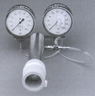
The actual maneuver depends somewhat on the equipment configuration. Respiratory pressures were originally measured using a pressure gauge and most early systems consisted of just a mouthpiece and a gauge (or gauges).

from 'Interpretation of Pulmonary Function Tests - A Practical Guide' by RE Hyatt, PD Scanlon and M Nakamura, Published by Lipincott-Raven, 1997, page 90.

from ‘Interpretation of Pulmonary Function Tests – A Practical Guide’ by RE Hyatt, PD Scanlon and M Nakamura, Published by Lipincott-Raven, 1997, page 90.

To use this type of system the patient either exhales to RV or inhales to TLC, places their lips around the mouthpiece and then forcefully inhales or exhales. Because of the limited amount of time available for lip placement a round plastic or cardboard mouthpiece is usually used.

Continue reading

Diagnosing Mitochondrial Myopathies

I’ve been reviewing my CPET textbooks trying to get a better idea of how to differentiate between different cardiovascular limitations. The other day I ran across an article on a related subject and thought it might be instructive.

The hallmark of cardiovascular limitations is the inability to deliver enough oxygenated blood to the exercising muscles. Another limitation that has similarities to this (and one that is infrequently diagnosed) is the inability of the exercising muscle to utilize the oxygen delivered to it. The best examples of this type of exercise limitation are mitochondrial myopathies (MM).

The mitochondria are the primary source of the ATP used by exercising muscle. There are several conditions that can cause the number of mitochondria to be reduced and there is wide variety of mitochondrial genetic defects. Mitochondria have their own genes and these can have both inherited or acquired genetic defects which can cause anything from mild to severe decreases in the ability to produce ATP. Mitochondria require oxygen to produce ATP so when the number of mitochondria are reduced or their ability to produce ATP is reduced the rate at which oxygen is consumed by an exercising muscle is also reduced.

A relatively common complaint of individuals with MM is dyspnea and exercise intolerance. One study found that 8.5% of all the patients referred to a dyspnea clinic had a mitochondrial myopathy of one kind or another. A definitive diagnosis requires a muscle biopsy but because the symptoms are often non-specific and a biopsy is an invasive procedure, it is usually not performed unless there more significant evidence suggesting a MM.

Continue reading

SNIP when you can’t MIP

The MIP test is used to assess the strength of the inspiratory muscles and is commonly performed in patients with neuromuscular disease. Results however, are often low because the sensation involved in performing a maximal inspiratory effort against an occluded airway is unpleasant and because the MIP requires coordination, cooperation and motivation to be performed correctly. In addition patients with neuromuscular disease frequently lack the muscular strength necessary to grip the mouthpiece and can therefore leak around it. For these reasons although a normal value will rule out significant muscular weakness, a low value can be difficult to interpret.

Sniff Nasal Inspiratory Pressure (SNIP) is an alternative way to measure inspiratory muscle strength that does not require a mouthpiece and uses a fairly natural maneuver. To perform the test one nostril is blocked with a soft probe attached to either a manometer or a transducer. The patient is asked to perform a normal exhalation to FRC and then inhale forcibly (sniff) with their mouth closed. The sniff effort is short since the maximum nasal inspiratory pressure is reached in a half a second or less.

Continue reading