When it’s FVC 1, EOT 2, volume comes out short

I was reviewing a pre- and post-bronchodilator spirometry report that showed a relatively large increase in FVC but the change in FEV1 was not significant. It’s not impossible for a patient to show this kind of a pattern following a bronchodilator but it is somewhat unusual. Usually when I see this it means that the patient exhaled a lot longer post-BD than they did pre-BD. When I looked however, I saw that just the opposite was true, the expiratory time was actually shorter for the post-BD effort than it was for the pre-BD effort.

FVC_Error_Table

The reported expiratory time isn’t always accurate, though. When a patient stops exhaling during an FVC effort but doesn’t inhale our test system will sometimes continue to time the effort. When this happens the volume-time curve becomes flat and the expiratory time is reported with a falsely high value.

FVC Early Termination

This is what I expected to see when I looked at the volume-time graphs for this report. What I saw instead was this:

Continue reading

Assessing the response to bronchodilators

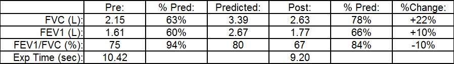
The current ATS/ERS guidelines require that an individual have a post-bronchodilator increase in FEV1 or FVC of at least 12% and 200 ml in order for it to be considered a significant response. Numerous studies have shown however, that many patients that don’t meet these criteria, particularly those with COPD, do have a clinically significant improvement with bronchodilators.

The September 2014 issue of Chest had a point-counterpoint set of editorials on this standard and on assessing the response to bronchodilator in general. Both sides had a number of interesting things to say but to a large extent one side was talking apples (physiology) and the other side oranges (statistics). I actually think that both sides feel there are significant problems with the ATS/ERS standards, they just differ in what they think is wrong and in the best way to fix it.

One statistical argument was that the ATS/ERS guideline is a one-size-fits-all solution that is designed more to detect asthmatic-type responses than the more subtle changes that can occur in individuals with COPD that although small, are clinically relevant. I am inclined to agree with this but as much as I and others think that the current ATS/ERS standard likely needs revision the difficulty with this is that spirometry is a “noisy” measurement with a lot of variability.

Spirometry variability and the limits to its accuracy comes from issues to familiar to all of us:

  • the patient’s ability to understand and perform spirometry
  • the technician’s ability to encourage and guide a patient through a spirometer maneuver
  • the spirometer’s ability to measure exhaled flow and volume accurately

“Noise” is one of the reasons why it can be difficult to determine statistically and clinically significant changes. It’s also why the threshold for significant post-bronchodilator change is set as high as it is and may also be the reason why it shouldn’t be lowered.

Even though there may be reasons why the ATS/ERS guidelines for post-bronchodilator improvements in FEV1 and FVC shouldn’t be revised at the very least they need to become more inclusive and consider more factors. There is too much emphasis placed on the changes in FEV1 and FVC as the sole indication for a response to bronchodilator particularly since there are other ways in which a response can be measured.

Continue reading

Is it Dynamic Hyperinflation or something else?

Patients with COPD often have a ventilatory limitation as their primary limitation to exercise. A ventilatory limitation to exercise has traditionally been assessed by the breathing index or the breathing reserve:

breathing index = Peak Ve / Predicted MVV

breathing reserve = 1 – (Peak Ve / Predicted MVV)

which are basically two different ways of saying the same thing. In either case a breathing index greater than 85% or breathing reserve less than 15% is an indication that a patient has reached a ventilatory limit to exercise. There is some disagreement as to whether the predicted MVV should come from a MBC test performed by the patient or from the patient’s FEV1 x 40. I have tried both approaches and my experience has been that FEV1 x 40 is the best indicator for a patient’s predicted MVV. This is also Wasserman’s (my go-to source for exercise testing) recommendation so this is what we use.

Individuals with COPD are occasionally hyperinflated at rest (i.e. elevated FRC and RV) and more commonly they dynamically hyperinflate during exercise. Research has shown that those individuals with are flow-limited during tidal breathing at rest almost always hyperinflate with exercise. Patients who are not flow-limited at rest but still have a low FEV1 and FEV1/FVC ratio may also hyperinflate. Because hyperinflation limits a patient’s tidal volume response to exercise it may cause an individual to have a limitation to exercise that occurs at a minute volume below the 85% threshold.

Exercise IC

Continue reading

Hidden FIVC

I was about to put a PFT report I’d been reviewing in my outbox when I noticed something odd about the flow-volume loop.

Hidden FIVC_redacted 1

What I saw was that the final inspiration of the FVC maneuver had ended to the left of the initial inspiration. This means a couple of thing, first and foremost that the FIVC was larger than the FVC and that the FVC was likely underestimated because the patient hadn’t really taken a full inspiration prior to exhaling. I had already looked at the raw data for the patient’s spirometry results for other reasons but I pulled them up again to see if I had missed something.

Continue reading