A change that probably isn’t a change

Recently a report came across my desk from a patient being seen in the Tracheomalacia Clinic. The clinic is jointly operated by Cardio-Thoracic Surgery and Interventional Pulmonology and among other things they stent airways. The patient had been stented several months ago and this was a follow-up visit. Given this I expected to see an improvement in spirometry, which had happened (not a given, BTW, some people’s airways do not tolerate stenting), but what I didn’t expect to see was a significant improvement in lung volumes and DLCO.

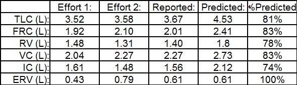
When I took a close look at the results however, it wasn’t clear to me that there really had been a change. Here’s the results from several months ago:

Observed: %Predicted: Predicted:
FVC: 1.19 50% 2.38
FEV1: 0.64 35% 1.79
FEV1/FVC: 53 71% 76
TLC: 3.21 76% 4.22
FRC: 2.34 96% 2.43
RV: 2.11 113% 1.85
RV/TLC: 66 150% 44
SVC: 1.15 48% 2.37
IC: 0.87 48% 1.80
ERV: 0.25 41% 0.58
DLCO: 6.59 38% 16.18
VA: 1.78 43% 4.12
IVC: 1.04

Change_that_isnt_change_2015_FVL_redacted_2

[more] Continue reading

Exercise and the IC, EELV and Vt/IC ratio

Determining whether a subject has a ventilatory limitation to exercise used to be fairly simple since it was based solely on the maximum minute ventilation (Ve) as a percent of predicted. There has been some mild controversy about how the predicted maximum ventilation is derived (FEV1 x 35, FEV1 x 40 or measured MVV) but these don’t affect the overall approach. Several decades ago however, it was realized that subjects with COPD tended to hyperinflate when their ventilation increased and that this hyperinflation could act to limit their maximum ventilation at levels below that predicted by minute ventilation alone.

The fact that FRC could change during exercise was hypothesized by numerous investigators but the ability to measure FRC under these conditions is technically difficult and this led to somewhat contradictory results. About 25 years ago it was realized that it wasn’t necessary to measure FRC, just the change in FRC and that this could be done with an Inspiratory Capacity (IC) measurement.

The maximum ventilatory capacity for any given individual is generally limited by their maximum flow-volume loop envelope. When a person with normal lungs exercises both their tidal volume and their inspiratory and expiratory flow rates increase.

Exercise_FVL_Normal

Exercise_VT_Normal

Continue reading

Is it Dynamic Hyperinflation or something else?

Patients with COPD often have a ventilatory limitation as their primary limitation to exercise. A ventilatory limitation to exercise has traditionally been assessed by the breathing index or the breathing reserve:

breathing index = Peak Ve / Predicted MVV

breathing reserve = 1 – (Peak Ve / Predicted MVV)

which are basically two different ways of saying the same thing. In either case a breathing index greater than 85% or breathing reserve less than 15% is an indication that a patient has reached a ventilatory limit to exercise. There is some disagreement as to whether the predicted MVV should come from a MBC test performed by the patient or from the patient’s FEV1 x 40. I have tried both approaches and my experience has been that FEV1 x 40 is the best indicator for a patient’s predicted MVV. This is also Wasserman’s (my go-to source for exercise testing) recommendation so this is what we use.

Individuals with COPD are occasionally hyperinflated at rest (i.e. elevated FRC and RV) and more commonly they dynamically hyperinflate during exercise. Research has shown that those individuals with are flow-limited during tidal breathing at rest almost always hyperinflate with exercise. Patients who are not flow-limited at rest but still have a low FEV1 and FEV1/FVC ratio may also hyperinflate. Because hyperinflation limits a patient’s tidal volume response to exercise it may cause an individual to have a limitation to exercise that occurs at a minute volume below the 85% threshold.

Exercise IC

Continue reading

COPD, BD and IC

The ATS/ERS recommendation for assessing the response to bronchodilator is based solely on changes in FEV1 or FVC. An FEV1 that does not improve significantly following bronchodilator inhalation is considered to be one of the hallmarks of COPD. Many individuals with COPD however, can have symptomatic relief and an improvement in their exercise capacity without a significant post-bronchodilator increase in FEV1. This means that FEV1 may not be the only criteria for assessing bronchodilator response.

One of the hallmarks of COPD is expiratory flow limitation. This can cause hyperinflation and is often reflected in an elevated FRC. It is also an important factor in exercise limitation. When ventilation increases during exercise in an individual with COPD, expiratory flow limitation causes the tidal volume and FRC to shift towards higher lung volumes. FRC is difficult to measure during exercise so this usually observed by measuring Inspiratory Capacity (IC).

IC Exercise COPD

COPD patients who don’t show a significant change in their FEV1 can respond to bronchodilators by becoming less expiratory flow-limited and when this happens their FRC decreases. Bronchodilator response in these individuals can therefore be assessed by measuring pre- and post-bronchodilator FRC or IC. At present there appears to be a consensus that an increase in IC or decrease in FRC of at least 0.30 liters or 12% should be considered to be a significant response.

Continue reading

An IC shows it’s probably not restriction

For the last couple of years it seems that I’ve had more problems than usual with lung volume tests. Even though this seems to date from the time that my lab went through its hardware and software upgrade and we started performing N2 washouts I suspect that these problems have been around for a long time and these events just heightened my awareness of lung volume testing problems.

My lab performs helium dilution, N2 washout and plethysmographic lung volume tests. When you are assessing the quality of lung volume tests the first problem for the helium dilution and plethysmographic techniques is whether or not the Functional Residual Capacity (FRC) was accurately measured and for N2 washout, it’s whether or not the Residual Volume (RV) was accurately measured. Leaks are always an issue for any of these measurement techniques and for helium dilution and N2 washout leaks will almost always cause the Total Lung Capacity (TLC) to be overestimated. For plethysmography the picture is less clear since leaks can cause TLC to be either over- or under-estimated.

Once you accept that the initial measurement of FRC or RV is accurate, however, the next question is whether the SVC is accurate or not. Since SVC is a more relaxed test than a forced vital capacity the SVC volume should be at least the same as the FVC volume and it is often larger. When I see an SVC that is smaller than the FVC I tend to think that the calculated TLC is probably okay and it’s the RV that is more likely to be overestimated. This is because the Inspiratory Capacity (IC) part of the SVC maneuver (“take as deep breath in as you can!”) is the easiest part and when the SVC is low, it is usually because the Expiratory Reserve Volume (ERV, “blow everything out that you can!”) is underestimated.

This report came across my desk a couple of days ago. The lung volumes were performed by helium dilution.

Not_RVD_Results 

Continue reading

The ERV Effect

I used to think that spirometry and diffusion capacity tests were hard and that lung volumes were easy. That may have been true in terms of getting patients to do the tests but I’ve long since come to the conclusion that it is easier to assess the quality of spirometry and diffusing capacity tests and know whether you have reasonably accurate results than it is to do this for lung volumes regardless of which lung volume measurement technique you use.

I was reviewing a set of plethysmographic lung volume tests when I noticed something very odd about the reported results. I usually look at just the VTG loops and the volume-time graphs in order to assess test quality. The testing software automatically selects and averages all VTG efforts and when I reviewed them there were a couple loops that were poor quality and I manually de-selected them. I was reviewing this report because the reported lung volume results didn’t quite match what the spirometry results were saying so this time I also took a close look at the numbers after I removed the low-quality loops. That’s when I realized that the reported TLC was larger than the two tests it was averaged from.

Pleth Math

Continue reading