Tag Archives: Fenyves & Gut

Spirometer, Pulmostar S, Fenyves & Gut, circa 1970

From a Fenyves & Gut sales brochure kindly provided by Emanuele Isnardi.

“Principle of measurement: the patient breathes room air (or, in special cases, a gas mixture) through a sterilisable, heated pneumotachograph head fitted to a mouthpiece of breathing mask. The resulting pressure different of a few millimetres water gauage picked up the the pneumotachotraph and proportional to the flow V is concerted to the corresponding positive and negative voltages by means of a pressure transducer and registered in an electronic integrator after amplification. The voltage resulting from this operation is proportional to the tidal volume (TV). The factor of the proportional relationship is determined by volumetric calibration, using a calibration pump. One litre of air is pumped through the pneumotachograph head, which is pre-heated to operating temperature. This is the only reasonable method of checking the entire system. A balance control compensates for any lack of symmetry between pneumotachograph heads.

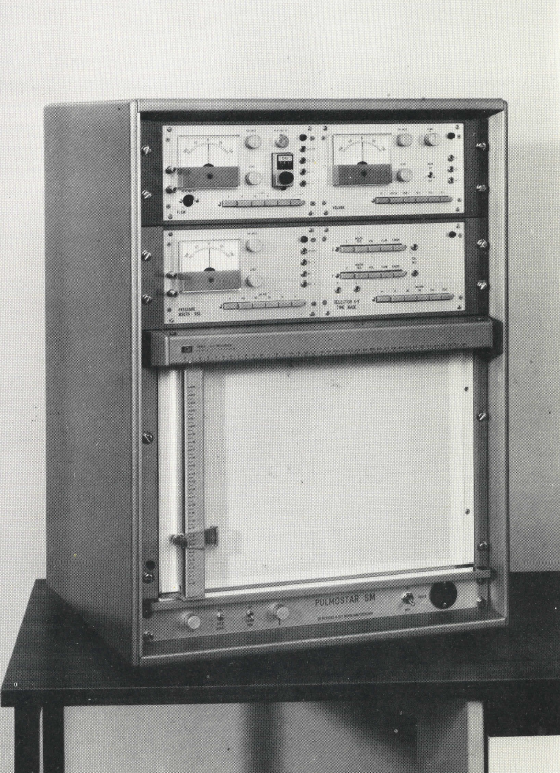
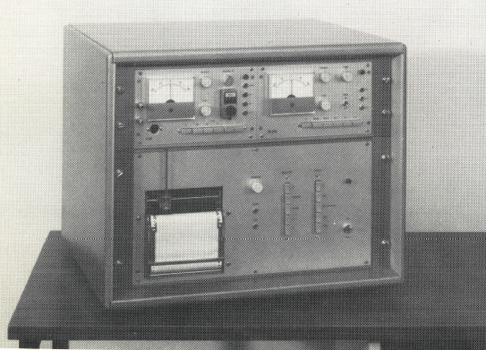
The S basic unit this consists of a slide-in unit for measuring the flow and a linked integrator and calibrating device. Measurements ranges: 1, 2, 5, 10, 20 litres or litres/sec., respectively, on full-scale deflection; time constants: 1000, 20 , 10, 5 seconds. Push-button selectors.  Automatic marking of zero-flow pointes V=0; heating of pneumotachograph head; data output using optional built-in single- or multi-channel compensated direct recordes or X-Y records; repectively.

Determinable parameters at rest and during exercise: tidal volume TV, inspiratory reserve volume IRV, expiratory reserve volume ERV, inspiratory capacity IC, forced inspiratory volume FIV, vital capacity VC, forced vital capacity FVC, forced expiratory volume FEV1, percentage expired %FEV1/VC and %FEV1/FVC, mean inspiratory flow MIFR, mean expiratory flow rate MEFR, mean flow rate MFR, peak inspiratory flow PIFR, peak expiratory flow rate PEFR, peak flow rate PFR, forced inspiratory flow FIF, forced expiratory FEF, forced mid-expiratory flow rate FMF, minute volume MV, maximum breathing capacity MBC, indirect maximum breathing capacity IMBC, maximum voluntary ventilation MVV, excess exercise ventilation EEV, standardised ventilation SV, ventilation in response to exercie VRE, respiratory frequency F, duration of inspiration, and expiration, respectively, flow-volume diagram also with He2 (X-Y recorder necessary).”

Metabolic system, PULMOSPORT, Fenyves & Gut, circa 1970

From a Fenyves & Gut sales brochure kindly provided by Emanuele Isnardi.

“One possible extension of the S basic unit is designed to determine the gas exchange or G or perform ergospirography, respectively, this representing a SG assembly. Unlike rival products this unit operates without valves, i.e. under optimum physiological conditions. For it should be noted that there are still systems on the market which require the subject to inhaled through a mask fitted with inspiration valves and exhale through a hose fitted to the mask. Of course such systems cannot work satisfactorily since they involve inspiration valves and because dead-space air is re-inhaled from the expiration hose. Our system has overcome these drawbacks.

Principle of measurement: the expired gases are continuously drawn off while changes in concentration levels occur. However, the average values necessary for further processing (e.g. O2 uptake) are drawn off in proportion to the expiratory flow rate by a special pump of our own design which can be modulated very rapidly. The expirate is then stored in a small collecting vessel. The resulting weighted samples are then analyzed in on-line analyzers for their O2 and CO2 concentrations (ΔFO2 and ΔFCO2 respectively).

Essentially the G apparatus consists of a slide-in unit for extraction proportionate to the flow of respiratory gases during expiratory phases. A device for averaging and converting the pulsating gas flow to a steady state required by the analyzers is included. The necessary equipment comprises O2 and CO2 and, optional, N2 analyzers (see under A), as well as a multi-channel compensated direct recorder (see under R). We recommend our slide-unit for determining the MV directly (unnecessarily if the assembly includes a computer) because it no only reduces working time, but also compensated for the time-lag occuring between the ΔFO2 and respectively,  ΔFCO2 quantities on the one hand, and the minute volume on the other.”

Exercise Analogue Computer, Fenyves & Gut, circa 1970

From a Fenyves & Gut sales brochure kindly provided by Emanuele Isnardi.

“We supply an economical analogue computer with permanent wiring for the automatic computation of parameters if immediate interest to the physician (MVBTPS, VO2STPD, VCO2STPD, RQ, respiratory equivalent RE, VO2/PR, f, caloric production). This computer which may be acquired in three stages processes all data provided by the S and G assemblies according to the exact formulae reduced to the usual gas conditions and taking into account atmospheric pressures and the time-lag between sensing ventilatory activity and the determination of gas exchange values.

Data output by printer and/or 12 channel point plotter.”

Douglas Bag Box, Fenyves & Gut, circa 1970

From a Fenyves & Gut sales brochure kindly provided by Emanuele Isnardi.

“Construction: A transparent plastic box on castors houses two bags containing a maximum of 100 litres each for the inspiratory and expiratory air respectively. A valve system connects the bags to a pneumotachograph which measures the inspiratory and expiratory air. A three-way stopcock makes it possible to let the patient breathe ambient air or air from the bag according to choice. The expirate can be collected in the expiratory bag or released to the atmosphere. Device for filling and fan for deflating the bags. The gas concentration can be measured in the bags and continually monitored at the mouth. Additional equipment: gas analyser, depending on test objectives (see under A), multi-channel compensated direct recorder (see under R), computer for direct MV determination.”

Respiratory Mechanics, SM assembly, Fenyves & Gut, circa 1970

From a Fenyves & Gut sales brochure kindly provided by Emanuele Isnardi.

“The S basic unit may be extended to a SM assembly by adding a slide-in unite for the measurement of oesphageal pressure, this making tests of respiratory mechanics possible. As the zero-flow points (i.e. the points of inversion) can be marked automatically, it is possible to determine the elastic axis in the diagrams of mouth volume in relation to oesaphageal pressure even under uncertain conditions (as they often occur in patients suffering from emphysema).  This makes evaluation considerably easier.”

Plethysmograph, PULMOSTAR SMB, Fenyves & Gut, circa 1970

From a sales brochure kindly provided by Emanuele Isnardi.  The entire sales brochure can be downloaded here.

“Our PULMOSTAR SMB is a whole-body plethysmograph of the constant-volume type, i.e. it measures pressure changes due to thoracic volume-change differences. Measurements are made against the pressure in a compensating chamber. The compensating vessel is built into the plethysmograph and communicating with it through a high resistance (time constants of 50, 10 and 20 seconds may be selected). This offsets slow, e.g. thermally induced pressure changes while the pressure fluctuations occurring simultaneously with respiration are registered. This also eliminates or dampens interfering influences reaching the plethysmograph chamber and the compensating vessel cophasically. The zero position can be restored at any time by allowing both the plethysmograph chamber and the compensator to assume atmospheric pressure through a valve system.”

Plethysmograph, PULMOREX SMB, Fenyves & Gut, circa 1970

From a Fenyves & Gut sales brochure kindly provided by Emanuele Isnardi.

“Our PULMOREX body plethysmograph incorporates the pressure-corrected flow principle, but also be equipped with the constant-volume mode of operateion in addition. Presently, this type of body plethysmograph is probably the most perfect.

In order to explain the function of this plethysmograph we will proceed from the simpler constant-pressure plethysmograph.  In 1960 Mead described his “volume displacement” plethysmograph which he used in conjunction with a Krogh spirometer to determine changes in chamber volume. We replaced the Krogh spirometer with a pneumotachograph and a linked integrator.  The resulting assembly is an “open-box” “volume-displacement”, i.e. constant-pressure plethysmograph.”