Plethysmographs are used to measure lung volume and airway resistance via Boyle’s Law. The basic concepts are simple but there are numerous variations. Although considered by most to be a modern invention its principles go well over 100 years.

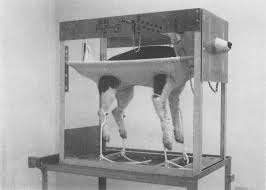
Animal Plethysmograph, 1891. From Physiologische graphik, By Oskar Langendorff, published by F. Deuticke , 1891, page 268. The plethysmograph is connected to a Marey tambour.
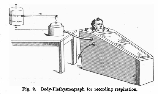
From Johns Scott Haldane and John Gillies Priestley. “The regulation of the lung‐ventilation.” The Journal of physiology 32.3-4 (1905): 225-266.
“In investigating the normal respiratory ventilation is is evidently important to avoid breathing through tubes, vales, etc,. and equally important to have a definitive quantitative record of the air breathed. In order to attain these two objects we employed a body plethysmograph arranged as follows (see Fig. 2).
“The box of the plethysmograph was of such a size and shape as to include as small a space as practicable, and yet enable the subject of the experiment to sit in perfect comfort. Its internal dimensions were as follows:–Floor, 45 inches long. Back, 19-1/2 inches wide x 24-1/4 inches. Lid, 19-1/2 inches wide behind x 16 inches in front x 14-3/4 inches. Sloping front, 37-1/4 inches x 16 inches x 10 inches. End, 10 inches wide by 12-1/2 inches high.”
“The lid was in two parts, with a circular aperature in the middle, through which the neck passed. Thumb-screws were provided for fastening it to the box, and all joints, screw-holes, etc., were made tight with “plasticene” (modelling wax). Air-tight connection was made between the neck of the subject and the lid of the box by means of a rubber collar with a broad flange, which was slipped over the head, and fitted the neck as tightly as possible without causing congestion. The flange was pressed down tightly on a layer of plasticene on the lid of the box by a ring of wood. This wooden ring was of course in two pieces, and was pressed down on the collar by means of thumb screws.
“In the side of the box were two holes into which tubes were fitted by means of corks. One of these was connected with a rubber tube for letting air into or out of the box to adjust the level of the writing-point and counteract, if necessary, the influence of rise of temperature in the box. The other was connected with the recording apparatus by means of rubber tubing 1 inch in diameter.
“The recording apparatus itself was arranged as shown in the figure. The cylincer suspended to the balance beam was of light tin plate. Two sizes were used — of 8 and 12 inches diameter — according to the depth of breathing expected. As the up and down movements of the writing-point traced on the smoked cylinder were proportional to those of the bell-jar.”
Plethysmograph, Boruttau, 1908. From: Lehrbuch der medizinischen Physik: Für Studierende und Ärzte zur Ergänzung jedes Lehrbuchs der Experimentalphysik by Heinrich Johannes Boruttau, published by Barth, 1908, page 131.
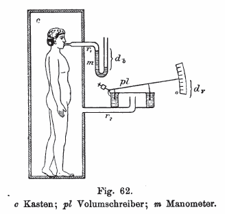
Plethysmograph, 1911. From Human Physiology, Volume 1, By Luigi Luciani, published by Macmillan and Company, 1911, page 422.
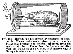
“The same effect may be obtained when the animal is breathing free air, while enclosed within a hermetically sealed glass cylinder (Knoll). A tube tied in the trachea, or fitting tightly over the mouth and nostrils of the animal, passes through the wall of one box and communicates with the external air. The internal air of the box is connected by means of a second tube with a recording tambour, and traces, like a plethysmograph, the variations in the total volume of the animal , corresponding to the inspiratory and expiratory movements. The simplest application of this method is that of Bernstein, represented in Fig. 188.
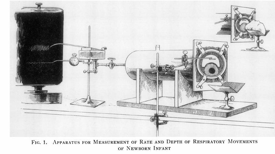
Infant Plethysmograph, 1931. From: J Clin Invest. 1931 August; 10(3): 545–558, BREATHING MEASUREMENTS ON NORMAL NEWBORN INFANTS, Douglas P. Murphy and Edward S. Thorpe, Jr.
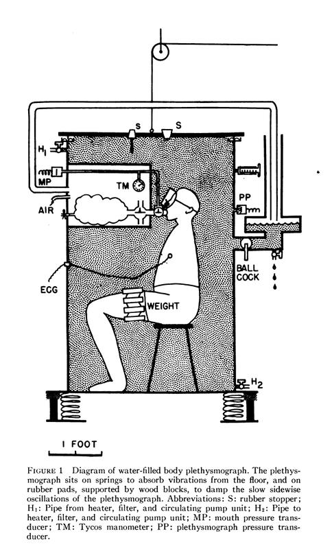
Water-Filled Plethysmograph, circa 1955. Found on the South Australia Medical Heritage website. Attributed to an article in the Journal of Clinical Investigations, vol 49, Issue 6 (June 1, 1970), pg 1238. Described as:
“The picture above shows a schematic design of one of DuBois’ early plethysmograph designs from the early 1950’s. The person sat inside a large box filled with warm water, which would allow for easy monitoring of volume changes. It is reported that as the person entered or left the box, it flooded the Dean’s office below. For some strange reason, this design did not last long.
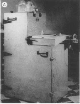
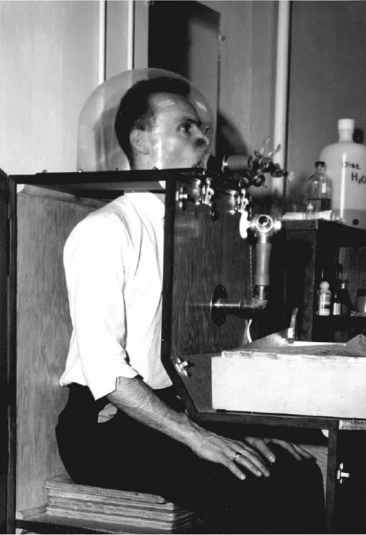
Volume Displacement Plethysmograph, 1960. Built by Jere Mead and J.H. Emerson. From Mead J. Volume displacement body plethysmograph for respiratory measurements in human subject. J Appl Physiol 1960; 15:736-740. Article provided by Carl O’Donnell, PhD, MPH
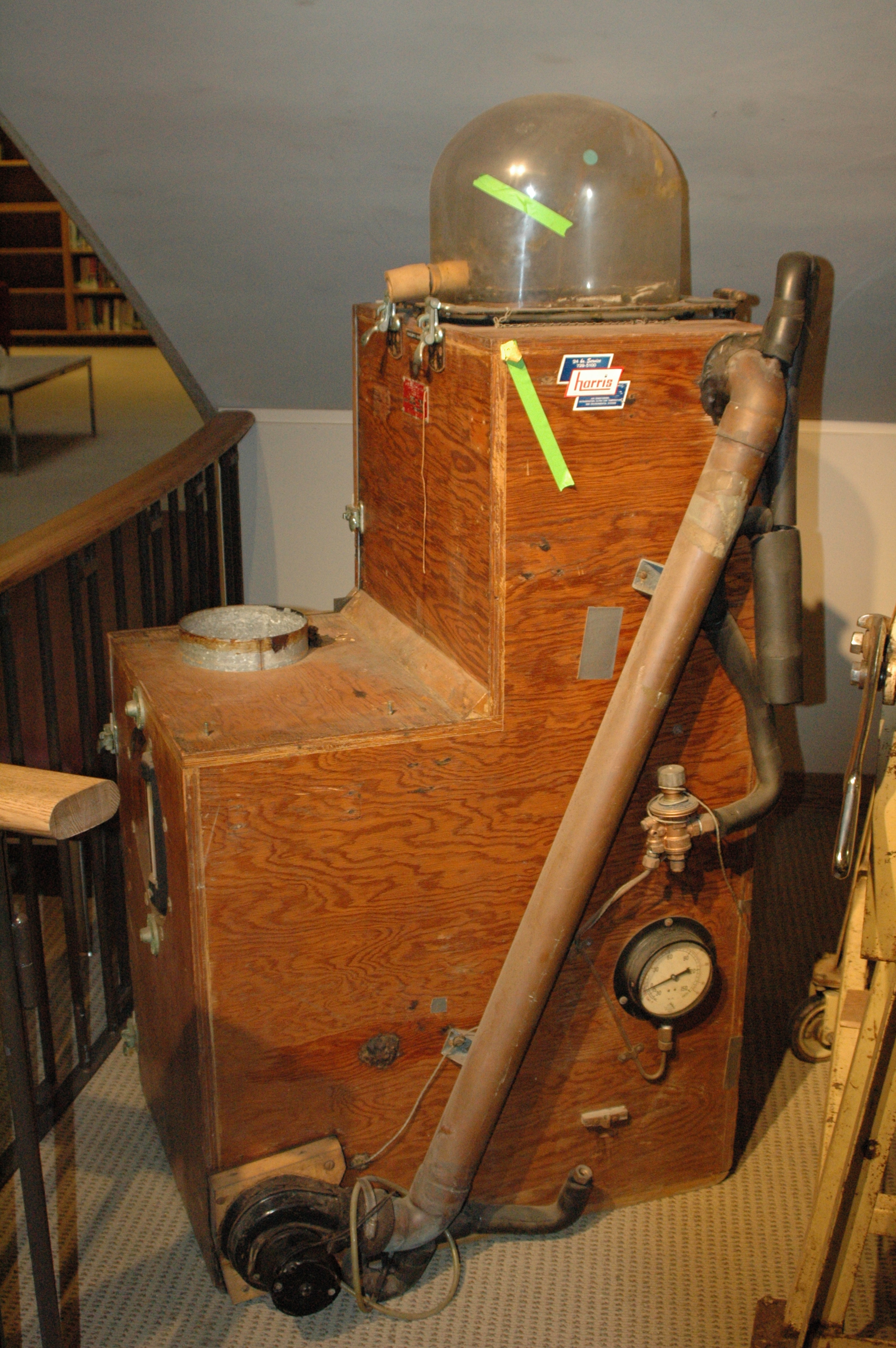
Volume Displacement Plethysmograph, 1960. Found on the Francis A. Countway Library of Medicine website. Warren Anatomical Museum: 21595. Experimental human body plethysmograph manufactured by JH Emerson Co. Machine was used to measure lung capacity. A person would sit inside the box with his head in the glass dome on top and breathe into the tube on front of dome. Machine is wooden and “L” shaped, designed to fit around a seated human, and latches closed. A spirometer would have rested on wooden edge. Two thermometers affixed to front measured interior and exterior temperatures. Tubing, gauge, and motor are affixed to right side. Rear of machine has attached wires and area marked “Sears-port”. 83 D x 59 W x 152 H cm.
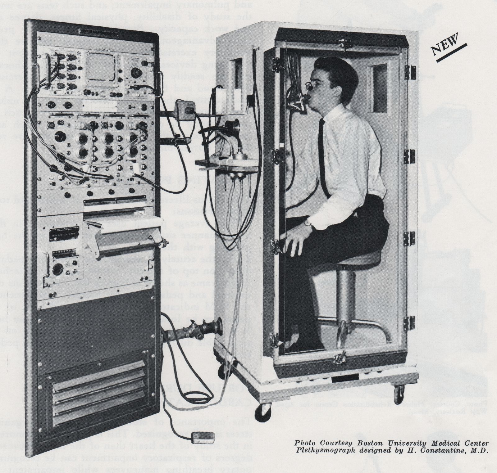
Plethysmograph, 1963. A constant pressure-variable volume plethysmograph that appears to have been based on the design of Mead and Emerson. From a doctoral thesis, “Acute effects of inhalation of cigarette smoke on pulmonary mechanics” by John M. Miller, University of Edinburgh, Edmonton, Canada, 1963, page 18.

Plethysmography, 1960’s. Found at Zeepreventorium.org. No description given. Probably from the 1960′s. It is likely a volume-displacement plethysmograph and was identified as being manufactured by Fenyves & Gut by Emanuele Isnardi.
Plethysmograph, Canine, 1964. From “A Canine Inhalation Exposure Apparatus Utilizing a Whole Body Plethysmograph.” by Boecker, B. B.; Aguilar, F. L.; Mercer, T. T., Health Physics, Volume 10, Issue 12, December, 1964.
Collins Plethysmograph, 1966. 1966 is the first year Collins sold a plethysmograph. From ‘A Catalog of Pulmonary Function Equipment and Accessories’, W. E. Collins Inc., 1966, page 49.

Med-Science Pulmo-Box Model 275, 1967. A volume-displacement plethysmograph. From Medical Electronic Laboratory Equipment 1967-68: Pergamon Electronics Data Series by G. W. A. Dummer, J. Mackenzie Robertson, published by Elsevier, page 826.
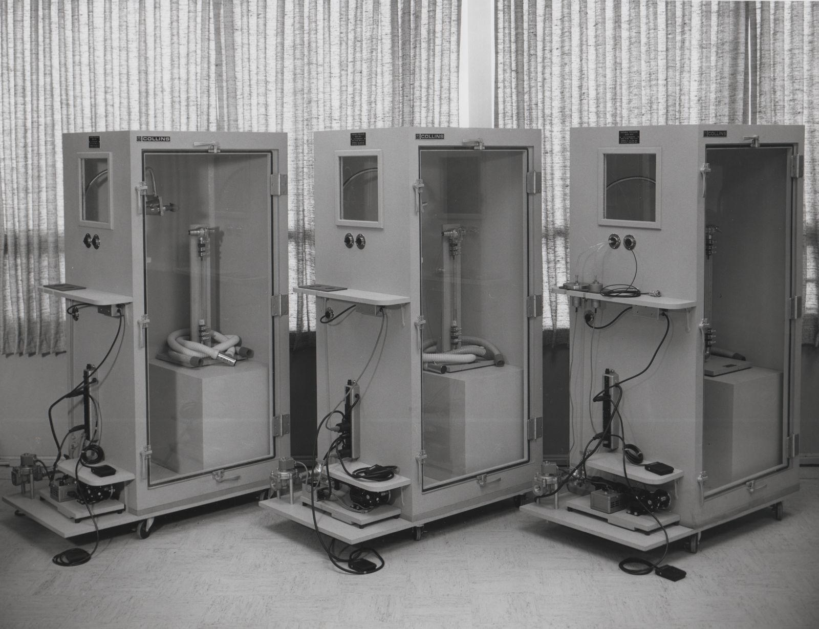
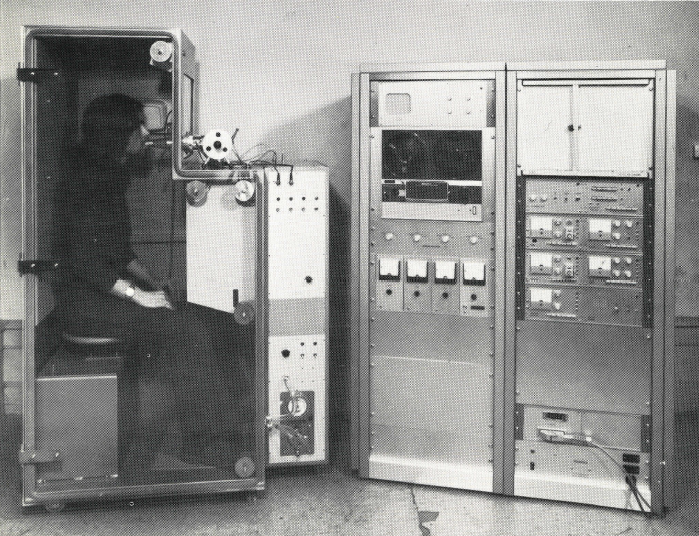
Collins Plethysmographs, circa 1968. From an undated publicity photo.
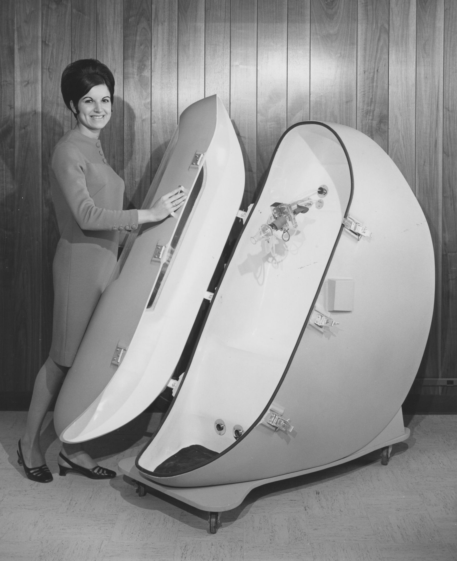
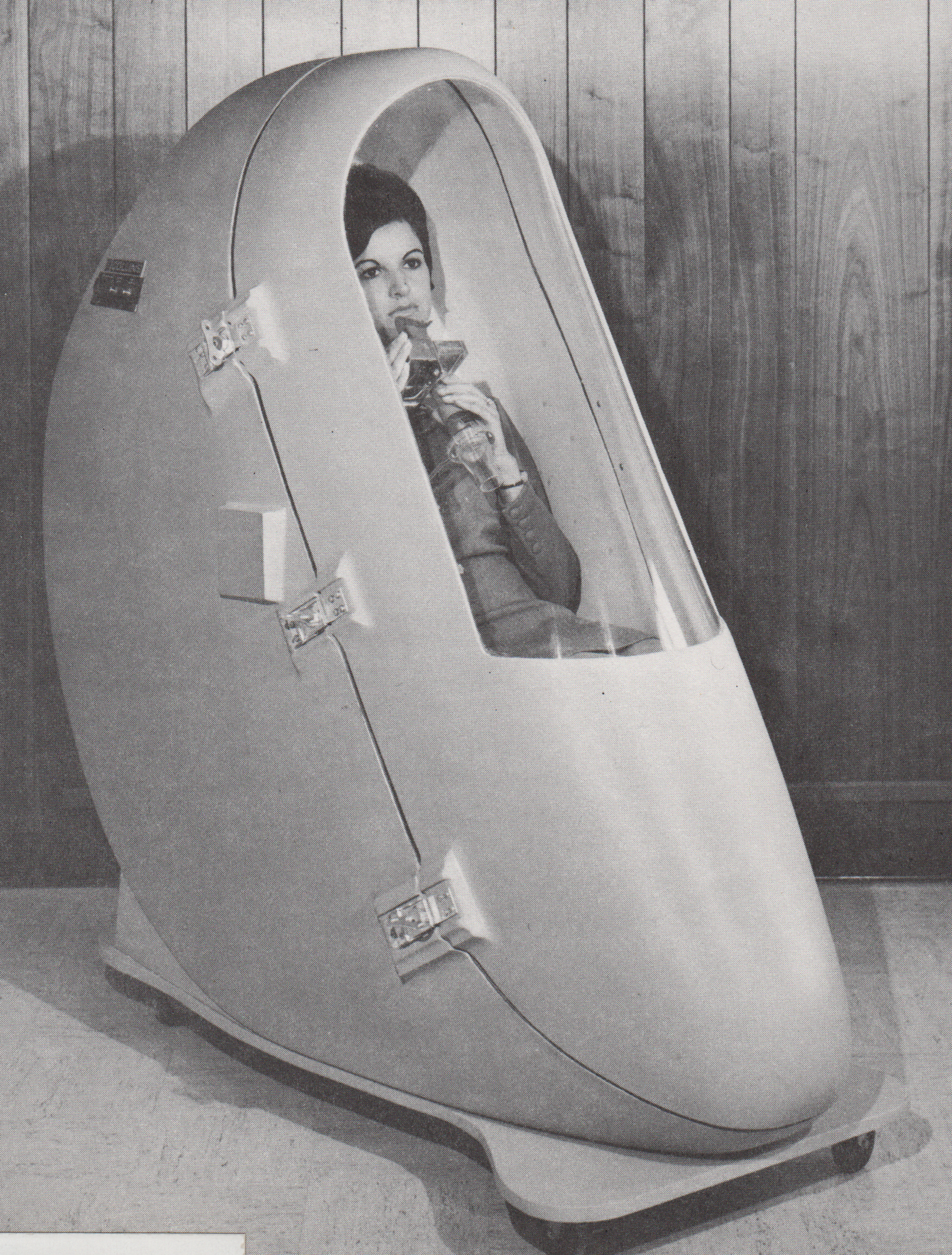
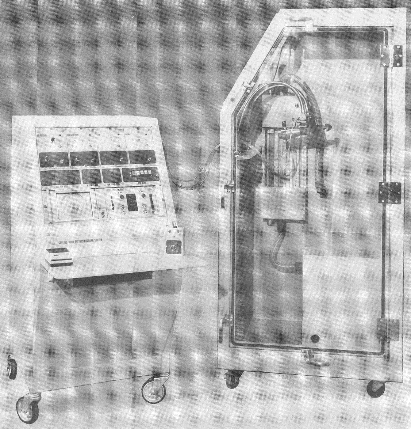
Fiberglass Plethysmograph, Collins, Model P-2506, 1969. From the 1969 Collins Equipment Catalog.
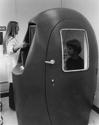
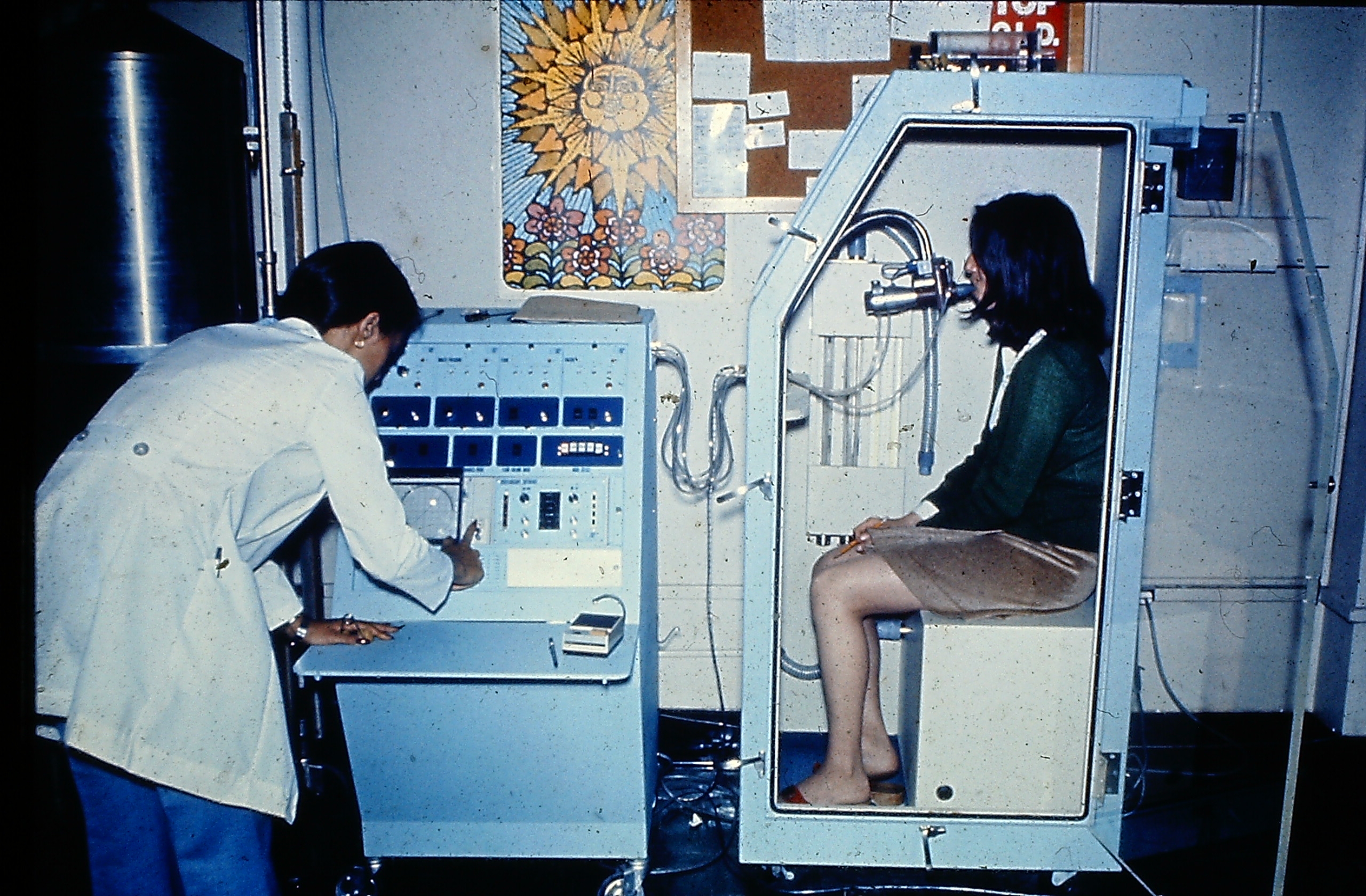
Plethysmograph, 1970’s. The manufacturer and exact date are unknown. Photo courtesy of James Sullivan, BA, RPFT, Supervisor, Pulmonary Laboratories, Memorial Sloan Kettering Cancer Center
Volume Displacement Plethysmograph, circa 1970’s. It was identified as being manufactured by Fenyves & Gut by Emanuele Isnardi. Found on an Autralian Health Service website.
Plethysmograph, Pulmostar SMB, Fenyves & Gut, circa 1970. From a sales brochure kindly provided by Emanuele Isnardi. The entire sales brochure can be downloaded here.
“Our PULMOSTAR SMB is a whole-body plethysmograph of the constant-volume type, i.e. it measures pressure changes due to thoracic volume-change differences. Measurements are made against the pressure in a compensating chamber. The compensating vessel is built into the plethysmograph and communicating with it through a high resistance (time constants of 50, 10 and 20 seconds may be selected). This offsets slow, e.g. thermally induced pressure changes while the pressure fluctuations occurring simultaneously with respiration are registered. This also eliminates or dampens interfering influences reaching the plethysmograph chamber and the compensating vessel cophasically. The zero position can be restored at any time by allowing both the plethysmograph chamber and the compensator to assume atmospheric pressure through a valve system.”
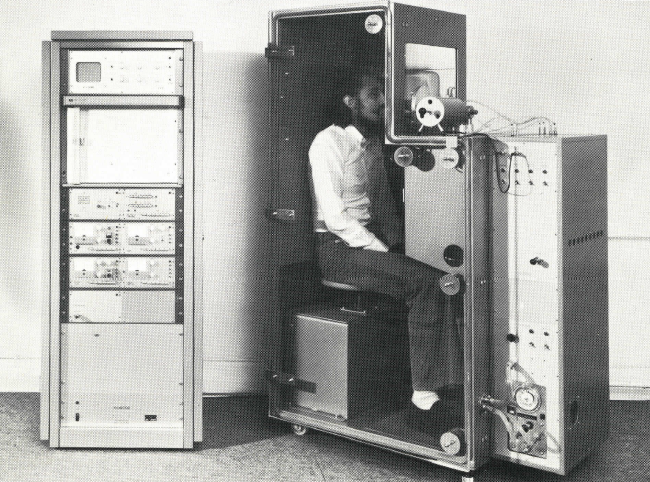
Plethysmograph, PULMOREX, Fenyves & Gut, circa 1970. From a sales brochure kindly provided by Emanuele Isnardi. The entire sales brochure can be downloaded here.
“Our PULMOREX body plethysmograph incorporates the pressure-corrected flow principle, but also be equipped with the constant-volume mode of operateion in addition. Presently, this type of body plethysmograph is probably the most perfect.
In order to explain the function of this plethysmograph we will proceed from the simpler constant-pressure plethysmograph. In 1960 Mead described his “volume displacement” plethysmograph which he used in conjunction with a Krogh spirometer to determine changes in chamber volume. We replaced the Krogh spirometer with a pneumotachograph and a linked integrator. The resulting assembly is an “open-box” “volume-displacement”, i.e. constant-pressure plethysmograph.”
Collins Plethysmograph, 1976. From Medical Instrumentation for Healthcare, by Cromwell L, Arditti M, Weibell FJ, Pfeiffer EA, Steele B, Labok J. Published by Prentice-Hall, 1976. Page 266.
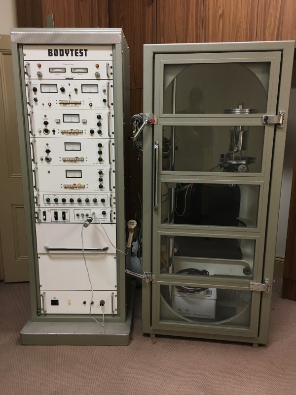
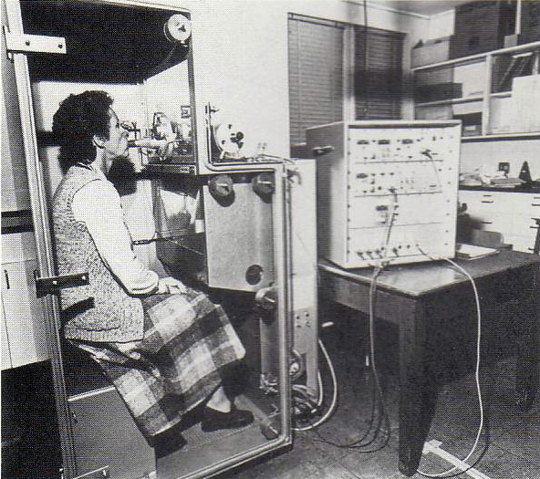
Jaeger Plethysmograph, 1978. A constant volume, variable pressure plethysmograph. From the South Australia Medical Heritage website. Described as:
“A Jäger BodyTest unit (left) with control machine (right). A DuBois type full body constant volume plethysmograph from c. 1978.”
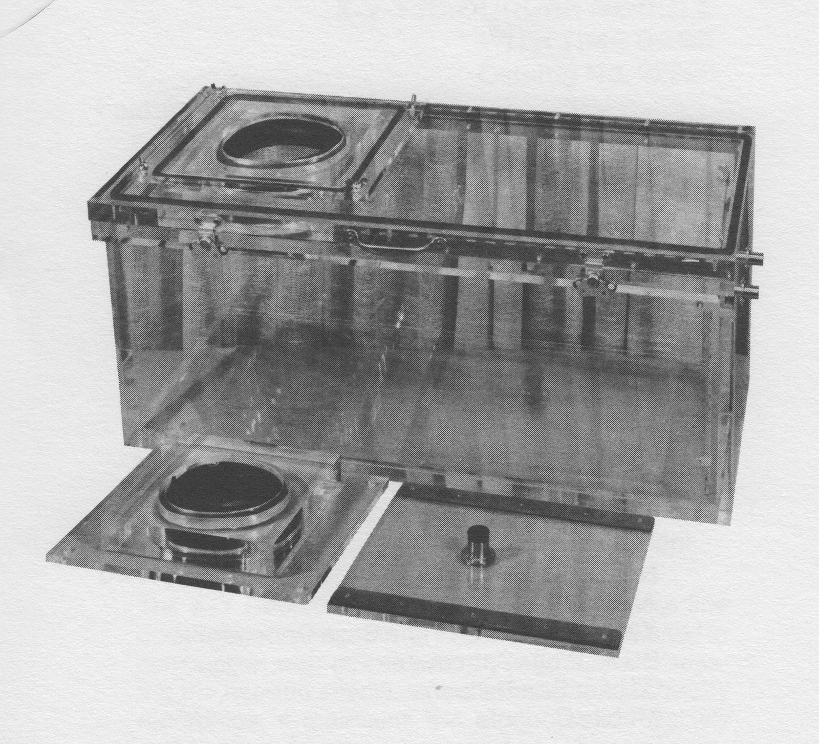
Collins Small Infant Plethysmograpy, circa 1978. Had to be custom ordered and you supplied your own pressure transducers. From ‘Collins’ Catalog of Pulmonary Function Testing Instruments”, undated by likely from around 1978, page 1-35.
Collins Plethysmograph, circa 1980. Photo courtesy of Carl O’Donnell, PhD, MPH.
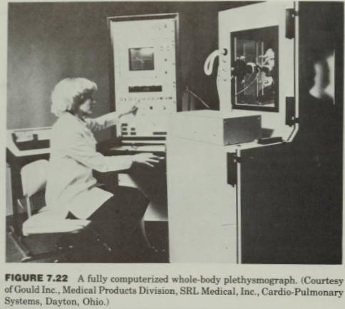
Gould Plethysmograph, 1982. From ‘Computer applications for patient care”, by Joseph D. Bronzino, published by Addison-Wesley, 1982, page 276.
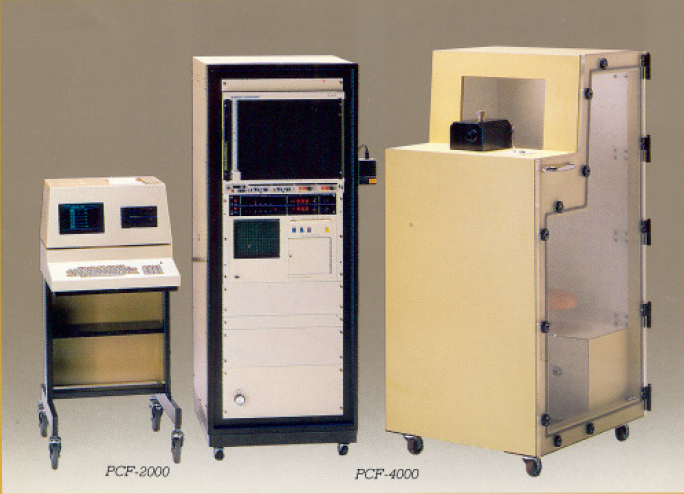
Fukuda Sangyo, PCF-4000, 1984: A transmural plethysmograph. The PCF-2000 was an optional digital computer for performing tests. Tests could be performed and calculated manually with just the PCF-4000.Photo from a sales brochure courtesy of James Sullivan, BA, RPFT, Supervisor, Pulmonary Laboratories, Memorial Sloan Kettering Cancer Center

Constant volume plethysmograph. Manufactured by Med Graphics in 1989. From a Pemed listing.
Collins Plethysmograph Box 1,1990. Photograph provided by Richard Johnston
Collins Infant Plethysmograph, circa 1995. Found at MedUsed.com.

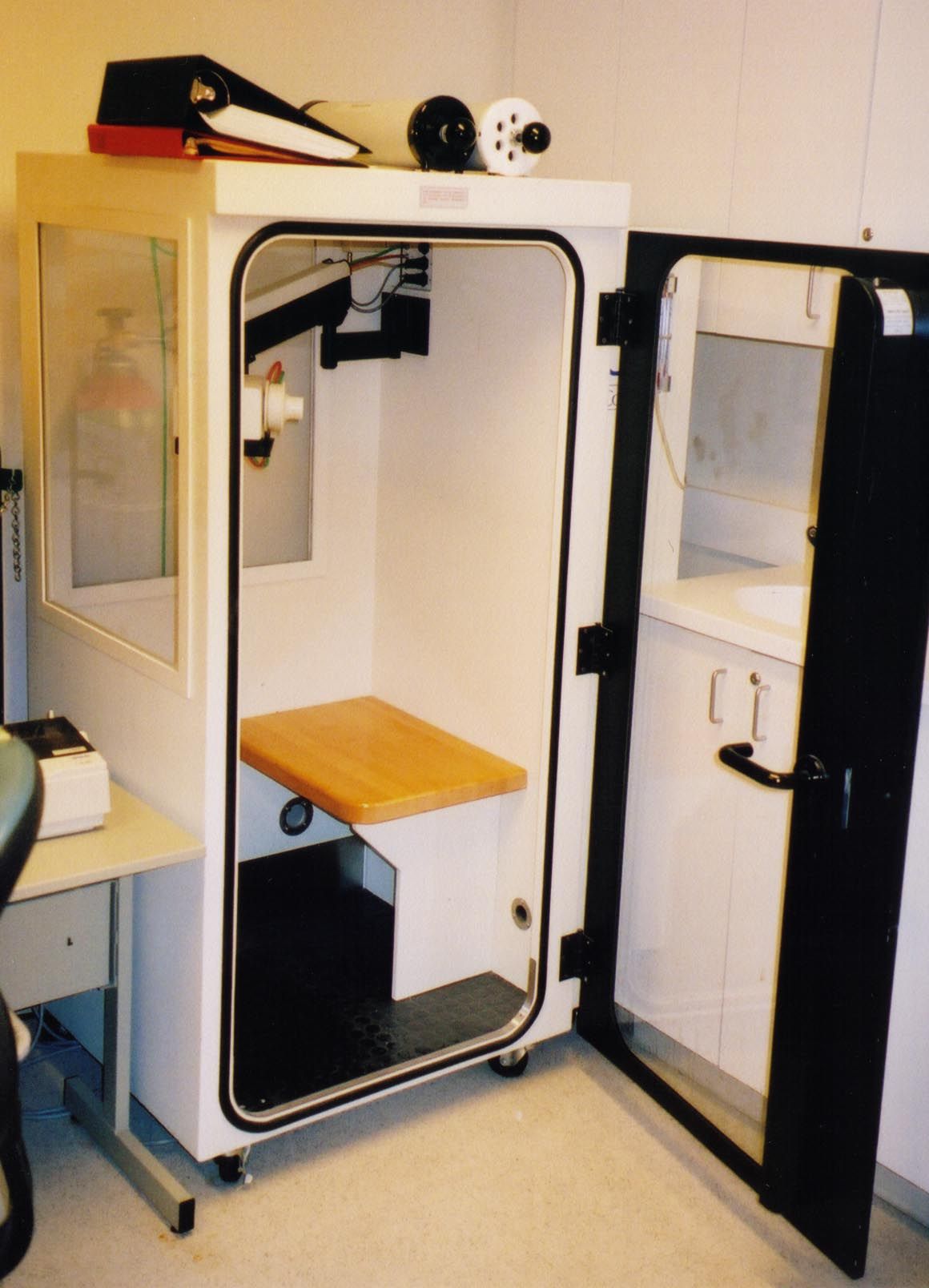
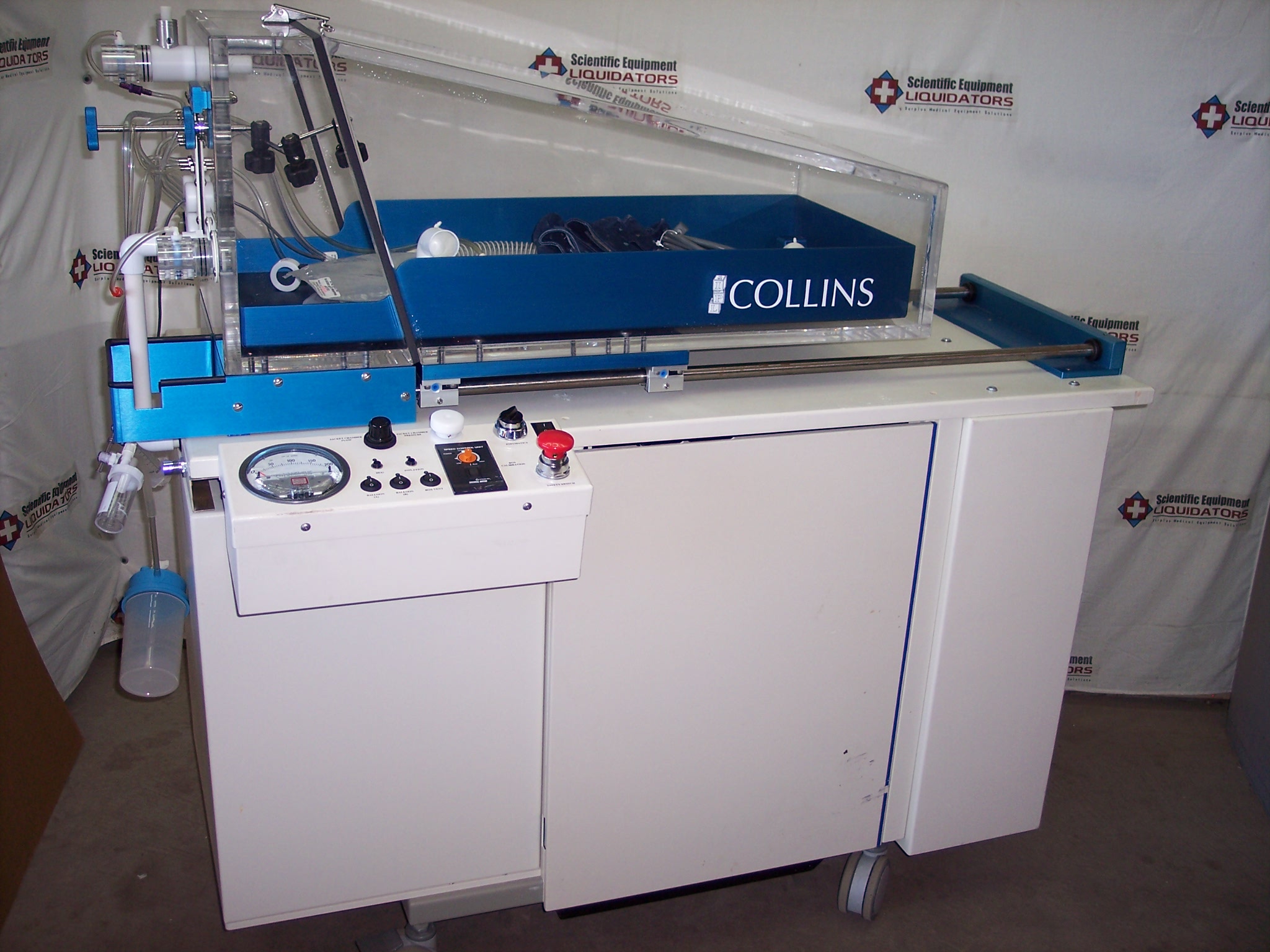
Sensormedics Constant Volume Plethysmograph, 2001. From a Pemed listing.
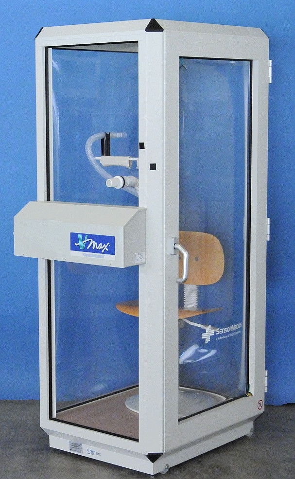
Sensormedics VMax V62J Autobox Plethysmograph, 2003. Found at a Pemed.com listing.
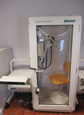
Jaeger Masterscreen Plethysmograph, circa 2005. From a DotMed listing.

PFT History by Richard Johnston is licensed under a Creative Commons Attribution-NonCommercial 4.0 International License.
Plethysmography, 1960’s and Volume Displacement Spirometer, circa 1970’s were not made by Godart or Minjhardt, but Fenjves & Gut. I worked with this unit until 1986. I have the instruction booklet.