From “Methods in pulmonary physiology”, by Bertels H, Bucherl E, Hertz CW, Rodewald G, Schwab M. Translated by Workman JM. Hafner Publishing Co., 1963, page 30. Undated, but probably from the mid-1930’s.
From “Methods in pulmonary physiology”, by Bertels H, Bucherl E, Hertz CW, Rodewald G, Schwab M. Translated by Workman JM. Hafner Publishing Co., 1963, page 30. Undated, but probably from the mid-1930’s.
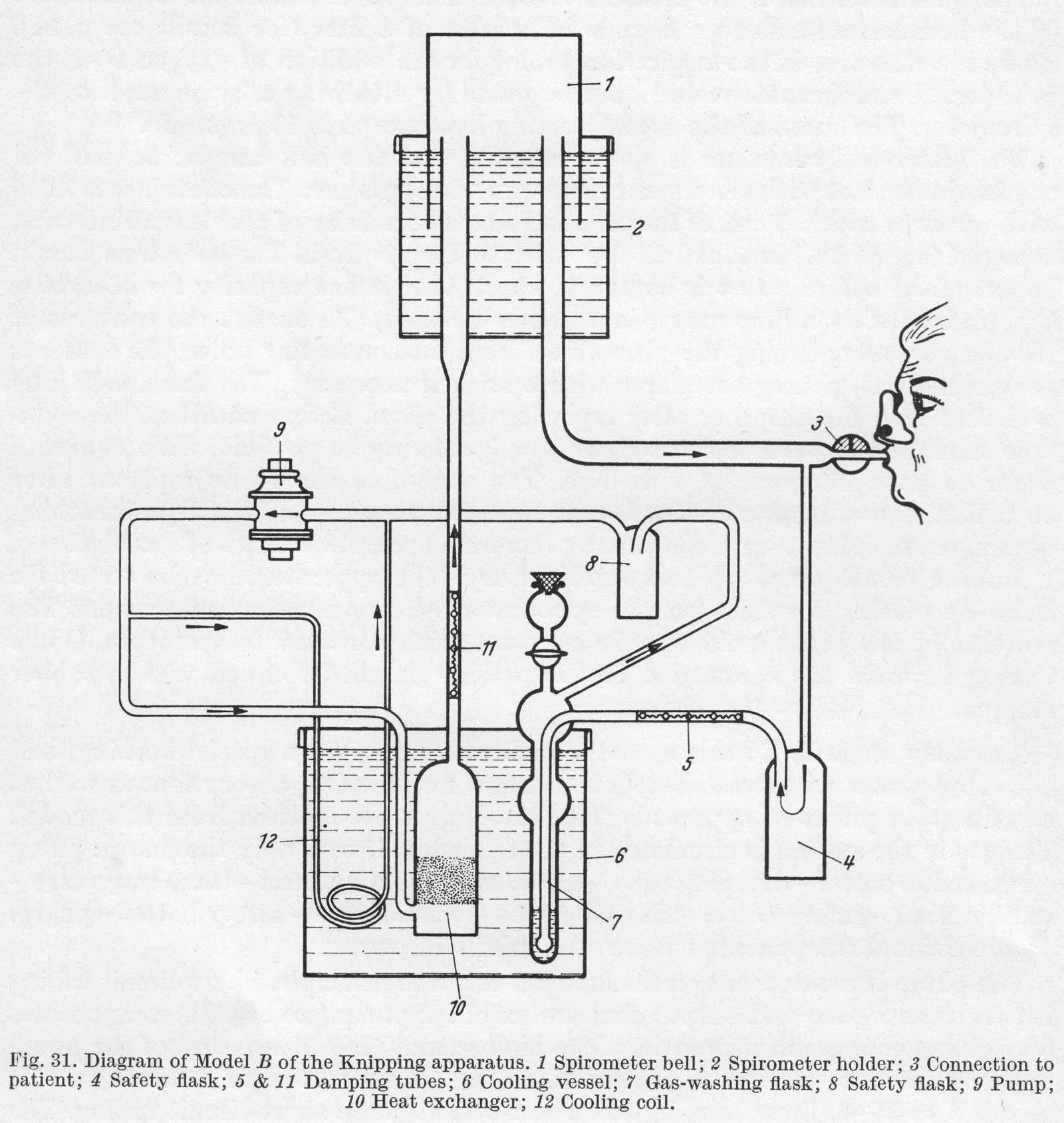
From “Methods in pulmonary physiology”, by Bertels H, Bucherl E, Hertz CW, Rodewald G, Schwab M. Translated by Workman JM. Hafner Publishing Co., 1963, page 26.
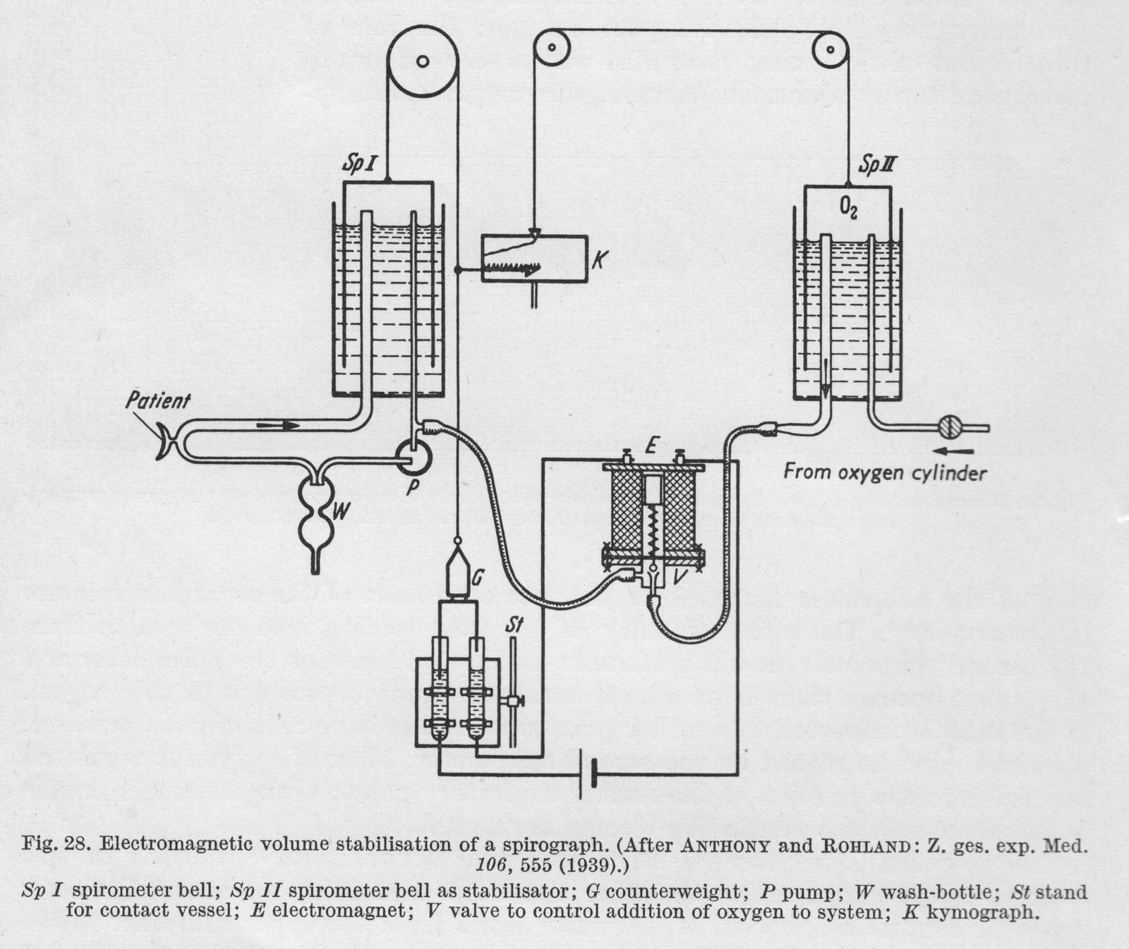
“Anthony and Rohland were the first to use an electro-magnetic device. In this apparatus, the addition of oxygen is dependent on the expiratory position. The counterweight of the spirometer bell dips into a vessel of mercury, so arranged that at the highest position of the spirometer (expiratory position) an electrical circuit is closed, resulting in the addition of oxygen through a valve operated by an electro-magnet. The loss in volume of the O2 gasometer (the second spirometer bell) is recorded providing a measure of the O2 consumption. The ventilation tracing is horizontal.”
From “Methods in pulmonary physiology”, by Bertels H, Bucherl E, Hertz CW, Rodewald G, Schwab M. Translated by Workman JM. Hafner Publishing Co., 1963, page 50.
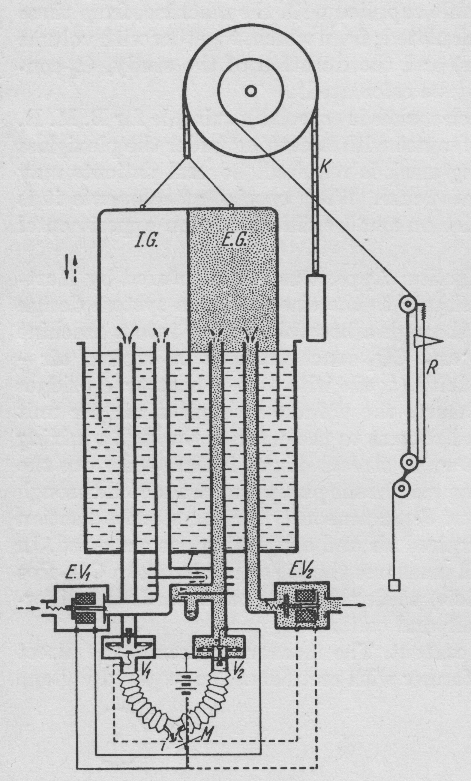
“An alternative method of making spirographic recordings from an open system by deloped by v. Tavel. A double spirometer is employed. The partition inside the spirometer bell separate the inspired from the expired air. The subject is connected to the apparatus by a mouthpiece or breathing mask. The course of the air is directed by valves V1 and V2. On inspiration air is withdrawn from the inspiratory section. On expiration air passes to the expiratory section. The tow controlling valves V1 and V2 control through corresponding contacts the opening and closing of the electromagnetic valves EV1 and EV2. On inspiration (when V1 is open) EV1 is closed and EV2 is opened. Thus during inspiration the bell sinks and air from IG enters the lungs, while the gas in EG passes through to the outside through EV2. On expiration (when V2 is open) EV1 us opened and EV2 is closed. This the bell rises during expiration because expired air passes into EG, and at the same time a corresponding volume of room air is drawn into IG through EV1. A system of coils T (heat exchanger) equalises the temperature of room air and expired air. The movements of the spirometer bell are recorded in the usual way on a kymograph. Since normally CO2 output is less than O2 uptake (RQ < 1), in this system there is a fall in the level of the bell which is proportional to the respiratory quotient (when RQ = 1, the course of the ventilation tracing is horizontal since the inspiratory and expiratory volumes are equal). Expired air may be collected for analysis in a Douglas bag attached distal to the valve EV2. Since the minute volume of ventilation is recorded on the kymograph and the respiratory quotient can be determined by the slope of the ventilation tracing, analysis of the CO2 concentration of expired air is all that is necessary for CO2 output and O2 uptake.”
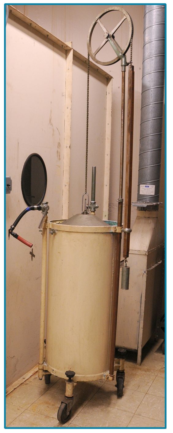
The age and manufacturer are unknown but the design is similar to Tissot spirometers manufactured between 1920 and 1950. Found on Pinterest.
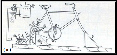
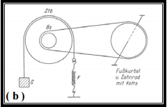
Bicycle ergometer developed by Holzer and Kalinka in 1935. It used friction from a metal strap around the wheel. Varying weights attached to the strap were used to adjust the workload. From a 2003 PhD dissertation by Yaser Mahfouz Atwa Saad Elgohari.
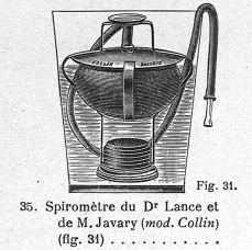
Spirometer attributed to Dr. Lance and M. Javary. Exactly how it worked is unclear. It may be a lung exercise device. From a Collin and Cie medical equipment catalog, Paris France, page 6. 1935. From the Biusante Website.
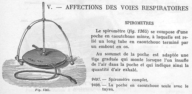
From a Rainal Brothers, Paris France, Medical Equipment Catalog, 1934, page 304. Found on the Biusante website.
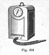
Probably a gas-meter spirometer. From the 1934 Medical Equipment catalog for H. Brodard, Paris, France. Page 53. Found on the Biusante website.
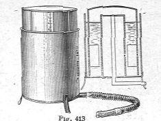
A water-seal spirometer. From the 1934 Medical Equipment catalog for H. Brodard, Paris, France. Page 53. Found on the Biusante website.
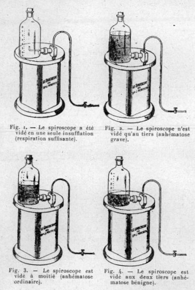
Dr. Pescher’s Spiroscope was primarily a device for exercising the lungs by moving water from the upper chamber to the lower. In this case, the amount of water that could be moved was used to diagnose the extent of a disease, most likely anemia from malaria. From Bulletins de la Société de pathologie exotique et de ses filiales de l’Ouest africain et de Madagascar, 1930, tome 23, page 138.