Making Assumptions about TGV and FRC

When lung volumes are measured in a plethysmograph the actual measurement is called the Thoracic Gas Volume (TGV). This is the volume of air in the lung at the time the shutter closes and the subject performs a panting maneuver. Ideally, the TGV measurement should be made at end-exhalation and should be approximately equal to the Functional Residual Capacity (FRC). For any number of reasons in both manual and automated systems this doesn’t happen and the point at which the TGV is measured is either above or below the FRC.

Testing software usually corrects for the difference in TGV and FRC by determining the end-exhalation baseline that is present during the tidal breathing at the beginning of the test. Using this value the software can determine where the TGV was measured relative to the tidal breathing FRC and then either subtracts or adds a correction factor to derive the actual FRC volume.

One problem with this is that leaks in either the subject or the mouthpiece and valve manifold can occur during the panting maneuver and the end-exhalation baseline can shift and this will affect the calculation of RV and TLC. I’ve discussed this previously and as a reminder, RV is calculated from:

RV = [average FRC] – [average ERV]

where the FRC is determined from the corrected TGV and ERV is determined from SVC maneuvers. TLC is then calculated from:

TLC = RV + [largest SVC]

When the post-shutter FRC baseline shifts upwards (higher lung volumes relative to the pre-shutter FRC):

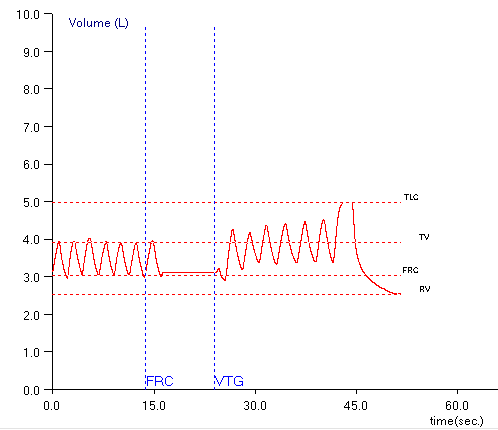
ERV is underestimated, which in turn causes both RV and TLC to be overestimated. When the post-shutter FRC baseline shifts downwards (lower lung volumes relative to the pre-shutter FRC):

ERV is overestimated, which in turn causes both RV and TLC to be underestimated.

I’ve been aware of this problem for quite a while and use this as a guideline when selecting the FRCs and SVCs from specific plethysmograph tests. All of these assumptions are based on the fact that FRC is derived from the pre-shutter end-exhalation tidal breathing. Well, you know what they say about assuming…

Continue reading

An unusual error in helium dilution lung volumes

Recently I was reviewing a report that included helium dilution lung volumes. What caught my eye was that the TLC and the FRC didn’t particularly fit in with the results from the other tests the patient had performed.

Test: Observed: %Predicted:
FVC: 2.83 114%
     
TLC: 3.03 71%
FRC: 0.88 39%
RV: 0.09 5%
SVC: 2.93 118%
     
VA: 3.64 88%

When compared to the FVC and the VA (from the DLCO test) the lung volumes are significantly lower. In particular the FRC and RV are markedly reduced. This is somewhat unusual for helium dilution lung volume since most errors usually cause FRC, RV and TLC to be over-estimated instead of being under-estimated. When I checked the other reports for the day I found that two other patients that had had their lung volumes measured on the same test system also had a TLC, FRC and RV that were noticeably reduced. Obviously we had some kind of equipment problem with that test system but it took a bit of sleuthing before I found out what had happened.

Like all lung volume tests, the helium dilution technique produces a lot of numbers, most of which are not included on the report. One of the first things I did was to call up the within-test data (our test systems store data every 15 seconds during the test and re-calculate FRC each time).

Time: FRC, Liters He conc. (%) Ve (L./min.) Vt, Liters
0:15 -1.00 9.71 6.16 0.21
0:30 0.06 8.87 10.1 0.59
0:45 0.43 8.61 11.76 0.78
1:00 0.69 8.44 9.05 0.72
1:15 0.76 8.39 8.18 0.74
1:30 0.79 8.37 8.32 0.59
1:45 0.82 8.36 8.15 0.62
2:00 0.83 8.35 7.79 0.65
2:15 0.86 8.33 5.51 0.62
2:30 0.87 8.32 5.34 0.63
2:45 0.88 8.32 0 0

When looking at this it was immediately evident there was a problem because the initial FRC was negative and this shouldn’t be possible. About the only way that helium dilution lung volumes can normally be underestimated is if the test is terminated way too early and the negative FRC ruled this out. It also narrows down the possible problems, but I had to think for a while and in doing so had to go back to the basics of the helium dilution test.

Helium dilution used to be the most common method for measuring lung volumes, but it requires a closed-circuit test system with a volume displacement spirometer. Most current test systems are open-circuit flow sensor-based systems and lung volumes are usually measured by nitrogen washout (or by plethysmography). Nevertheless, there are a couple of closed-circuit systems still being manufactured and there are a fair number of these systems still in service.

Continue reading

Exercise and the IC, EELV and Vt/IC ratio

Determining whether a subject has a ventilatory limitation to exercise used to be fairly simple since it was based solely on the maximum minute ventilation (Ve) as a percent of predicted. There has been some mild controversy about how the predicted maximum ventilation is derived (FEV1 x 35, FEV1 x 40 or measured MVV) but these don’t affect the overall approach. Several decades ago however, it was realized that subjects with COPD tended to hyperinflate when their ventilation increased and that this hyperinflation could act to limit their maximum ventilation at levels below that predicted by minute ventilation alone.

The fact that FRC could change during exercise was hypothesized by numerous investigators but the ability to measure FRC under these conditions is technically difficult and this led to somewhat contradictory results. About 25 years ago it was realized that it wasn’t necessary to measure FRC, just the change in FRC and that this could be done with an Inspiratory Capacity (IC) measurement.

The maximum ventilatory capacity for any given individual is generally limited by their maximum flow-volume loop envelope. When a person with normal lungs exercises both their tidal volume and their inspiratory and expiratory flow rates increase.

Exercise_FVL_Normal

Exercise_VT_Normal

Continue reading

COPD, BD and IC

The ATS/ERS recommendation for assessing the response to bronchodilator is based solely on changes in FEV1 or FVC. An FEV1 that does not improve significantly following bronchodilator inhalation is considered to be one of the hallmarks of COPD. Many individuals with COPD however, can have symptomatic relief and an improvement in their exercise capacity without a significant post-bronchodilator increase in FEV1. This means that FEV1 may not be the only criteria for assessing bronchodilator response.

One of the hallmarks of COPD is expiratory flow limitation. This can cause hyperinflation and is often reflected in an elevated FRC. It is also an important factor in exercise limitation. When ventilation increases during exercise in an individual with COPD, expiratory flow limitation causes the tidal volume and FRC to shift towards higher lung volumes. FRC is difficult to measure during exercise so this usually observed by measuring Inspiratory Capacity (IC).

IC Exercise COPD

COPD patients who don’t show a significant change in their FEV1 can respond to bronchodilators by becoming less expiratory flow-limited and when this happens their FRC decreases. Bronchodilator response in these individuals can therefore be assessed by measuring pre- and post-bronchodilator FRC or IC. At present there appears to be a consensus that an increase in IC or decrease in FRC of at least 0.30 liters or 12% should be considered to be a significant response.

Continue reading

What’s abnormal about FRC?

I’ve had a number of reports across my desk in the last couple of weeks with both elevated and reduced FRC’s that were associated with a more-or-less normal TLC. I reviewed the raw data from all of these tests (I review the raw data from all lung volume tests) and in only a few instances did I make any corrections to the report. This made me think however, about what, if anything, is an abnormal FRC trying to tell us?

The answers to that question range from “a whole bunch” to “not much” to “darned if I know”. When you measure lung volumes TLC is really the only clinically important result. RV can be useful at times but although the other lung volume subdivisions may play a role in the measurement process they have only a limited diagnostic value. All lung volume measurements start with FRC, however, and if you don’t know you have an accurate FRC how do you know that TLC is accurate?

FRC is a balance point of opposing forces in the lung and thorax. Lung tissue wants to collapse, the rib cage wants to spring open and the diaphragm wants to do whatever muscle tone, gravity and the abdomen allows it to do. All of these forces are to one extent or another dynamic and can change over time. These changes can occur both slowly and rapidly, and are the primary reason why isolated changes in FRC don’t tend to have a lot of clinical significance. For all lung volume measurements however, one primary assumption is that FRC does not change during the test and this isn’t necessarily true.

Continue reading