Ventilatory Challenge Testing

Airway hyper-responsiveness is a primary feature of asthma. There are a number of bronchial challenge tests designed to evoke and measure this factor, the most common of which require the inhalation of one or another bronchoconstrictive agent such as methacholine, histamine, mannitol or hypertonic saline.

An elevated ventilation can cause many asthmatics to bronchoconstrict and this is often the cause of Exercise-Induced Bronchospasm (EIB). There are two competing theories as to why this happens. A number of researchers have suggested that the mechanism is a drying of the airway mucosa which changes the osmolarity of the respiratory tract fluid which in turn causes some cells to releases mediators that cause bronchoconstriction. Other researchers assert that it is the cooling of the airways during hyperventilation and an increased blood flow and edema during subsequent re-warming that causes the bronchoconstriction. There is evidence to support both interpretations and it is likely that both mechanisms coexist, with one or the other being more predominant in any given individual.

Although the inhalation challenge tests are reasonably sensitive not all patients with EIB have a positive reaction. When a patient’s primary complaint is exercise-related or when they have had a negative inhalation challenge test and are still symptomatic, a ventilatory challenge test should be considered. There are several ventilatory challenge tests that are specifically oriented towards evoking and characterizing EIB. These are the Cold Air challenge, Eucapnic Voluntary Hyperventilation and Exercise Challenge. There are a number of similarities between these tests.

Cold Air Challenge

A Cold Air Challenge (CACh) test consists of having a patient hyperventilate while breathing air that has been cooled to a temperature of between -10°C and -20°C. It is usually performed using a mixture of 5% CO2, 21% O2, 74% N2 in order to prevent dizziness from hypocapnia.

Continue reading

The CPET’s not over until it’s over

My guidelines for interpreting CPETs originally started as notes to myself about what needed to be on the report and what the normal values were. They grew into a more formal set of instructions that were given to the pulmonary fellows when they were reviewing CPETs. Lately I’ve been reviewing them and re-reading the source material in order to make sure what I had written was still correct and so I could add references.

I tend to focus on one aspect of testing at a time and noticed while reviewing material that the measurements made at peak exercise or anaerobic threshold are almost always considered to be the most important test values. This is true to an extent, but information gathered both before and after testing is also important. In particular, after exercise has ended, during the recovery period, there are several measurements that should be made routinely and are diagnostically significant.

Heart rate recovery (HRR)

When exercise ends, an individual’s heart rate should start decreasing from its peak value. The heart rate recovery (HRR) is the difference between the heart rate at peak exercise and at some interval, usually 1 minute, post-exercise. The rise in heart rate during exercise is due to a combination of parasympathetic withdrawal and sympathetic activation. Vagal reactivation is the principle determinant of heart rate recovery after exercise and a reduction in the heart rate recovery (HRR) indicates a decrease in vagal tone specifically, and parasympathetic tone generally.

Continue reading

What does it mean when Ve exceeds its predicted during a CPET?

When I review the results from a CPET I am used to considering a maximum minute ventilation (Ve) greater than 85% of predicted as an indication of a pulmonary mechanical limitation. Recently a CPET report came across my desk with a maximum minute ventilation that was 142% of predicted. How is this possible and does it indicate a pulmonary mechanical limitation or not?

It is unusual to see a Ve that is greater than 100% of predicted. We derive our predicted max Ve from baseline spirometry and calculate it using FEV1 x 40. We have tried performing pre-exercise MVV tests in the past and using the maximum observed MVV as the predicted maximum Ve but our experience with this has been poor. Patients often have difficulty performing the MVV test correctly and realistically even if it is performed well the breathing maneuver used during an MVV test is not the same as what occurs during exercise. Since both Wasserman and the ATS/ACCP statement on cardiopulmonary exercise testing recommend the use of FEV1 x 35 or FEV1 x 40 as the predicted maximum minute ventilation we no longer use the MVV.

There are usually only two situations where a patient’s exercise Ve is greater than their predicted max Ve. First, when a patient is severely obstructed their FEV1 is quite low and FEV1 x 40 may underestimate what they are capable of since they are occasionally able to reach a Ve a couple of liters per minute higher than we expected. Second, if the FEV1 is underestimated due to poor test quality then the predicted max Ve will also be underestimated. In this case however, the baseline spirometry had good quality, was repeatable and the results did not show severe obstruction but instead looked more like mild restriction.

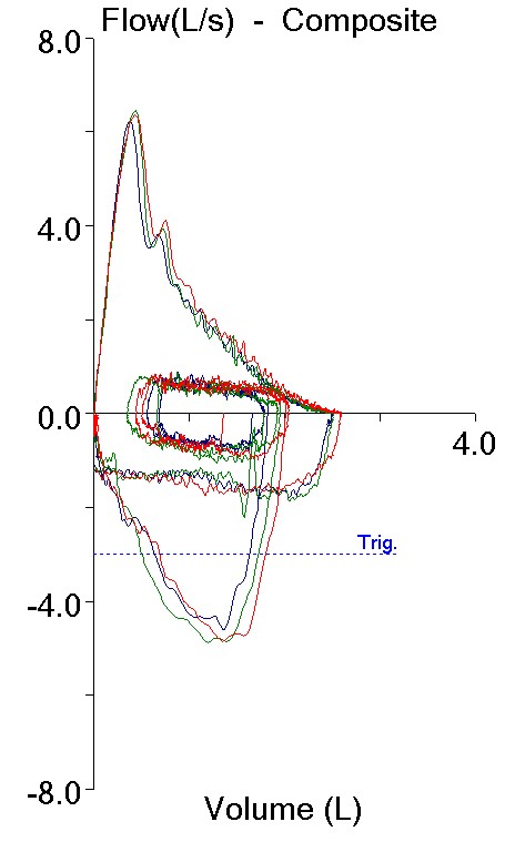
Pre_Exercise_FVLs

Effort 1: Effort 2: Effort 3:
FVC (L): 2.51 2.52 2.60
FEV1 (L): 1.86 1.87 1.95
FEV1/FVC %: 74 74 75
PEF: 6.26 6.46 6.37

Continue reading