Airway obstruction and the FVC

Spirometry is the most commonly performed (and mis-performed) pulmonary function test around the world. The apparent simplicity of spirometry is misleading since there are numerous subtleties that have a significant effect on the results.

I suspect that when the FVC is thought about it is most often considered to be an index towards the total capacity of the lung. That’s certainly true in it’s own way, but the FVC is actually a critically important factor when determining airway obstruction. I’ve had a number of reports across my desk lately where the patient had a reasonably large change in FVC when compared to their last visit but little change in FEV1, and this has made a difference in how the results are interpreted. For example:

Visit 1: Observed: %Predicted: Predicted:
FVC: 4.27 87% 4.91
FEV1: 3.36 84% 3.99
FEV1/FVC: 79 96% 82
Visit 2: Observed: %Predicted: Predicted:
FVC: 4.67 95% 4.91
FEV1: 3.38 85% 3.99
FEV1/FVC: 72 88% 82

Although the change in FVC is not significant by my lab’s standards (+0.40 L, +9%) and the FEV1 has hardly changed at all, the FEV1/FVC ratio has gone from being within normal limits to being under the LLN and therefore showing mild airway obstruction. Continue reading

Underutilized spirometry, missed opportunities

A friend is taking her father to a PFT lab (2500 miles away from where I am the moment so I couldn’t go along with them) because he has been short of breath for a couple of years, but oddly enough, only when lying on his side. I expect that despite these rather specific symptoms he will only get routine spirometry. I don’t necessarily fault the PFT Lab he’s going to for this, partly because physician orders often don’t include specifics, partly because they may not have the facilities to perform supine or lateral spirometry, and partly because its not clear lateral spirometry would show anything.

I don’t think that my lab is necessarily any better. We have only one room with an exam table that allows us to perform positional spirometry and that is largely because of the ALS patients we regularly see. Even so, unless we received specific orders to perform supine or lateral spirometry it’s unlikely that one of our technicians would think it was necessary and then take it on themselves to perform it. That itself is part of the problem not only for my lab but for the field of Pulmonary Function testing in general (but that’s another story).

The real problem however, is that the way in which spirometry is performed around the world is focused almost exclusively on detecting expiratory airway obstruction. It may be true that airway obstruction is primarily expiratory, but this ignores that fraction of individuals who have some degree of inspiratory obstruction. It also overlooks those individuals whose FVC is underestimated and FEV1/FVC ratio overestimated due to some degree of gas trapping. It also overlooks individuals that have positional airway obstruction that is not evident in the upright position.

We’ve fallen into the trap of thinking that there’s only one way to perform spirometry, and this is a mistake.

Continue reading

RVD’s and OVD’s can’t mix without the FEV1/FVC ratio

The patients whose reports I review have always been very accommodating. An issue of one kind or another catches my attention and before I know it I find several more reports that are similarly involved. Thanks to our patients I’ve had a number of reports come across my desk recently that showed a combination of restrictive and obstructive defects. This particular one may not be the best possible example but it seems to illustrate several points fairly well.

Observed: %Predicted: Predicted:
FVC (L): 1.12 40% 2.80
FEV1 (L): 0.75 35% 2.16
FEV1/FVC (%): 67 86% 78
TLC (L): 1.92 42% 4.54
FRC (L): 1.18 48% 2.47
RV (L): 0.76 44% 1.73
RV/TLC (%): 40 104% 38

Interpreting results like this as combined (or mixed) defects using the ATS/ERS algorithm seems relatively straightforward.

ATS-ERS Algorithm 2

From Brusasco V, Crapo R, Viegi G. ATS/ERS Task Force: Standardisation of pulmonary function testing. Interpretive strategies for lung function tests. Eur Respir J 2005; 26, page 956

The algorithm starts by using the FEV1/FVC ratio to determine whether obstruction is present and only then considers whether or not the FVC and TLC are normal. It occurred to me however, that this assumes that the normal range of the FEV1/FVC ratio is preserved when TLC decreases below normal. Given the markedly different causes of restrictive lung disease it would seem that saying that the FEV1/FVC ratio should remain within the normal range over a relatively broad range of lung capacities (and without necessarily knowing the cause for any reduction) seems a bit far-fetched. Interestingly enough however, it actually turns out to be reasonably true.

Continue reading

The NHANESIII FEV1/FVC ratio and height, revisited

I was reading James Hansen’s textbook on pulmonary function testing and ran across a spot where he made a minor criticism of the NHANESIII (Hankinson et al) reference equations for the FEV1/FVC ratio. Specifically he noted that the NHANESIII equation ignored height and only used age as a variable but that when he compared the directly calculated FEV1/FVC ratio with one indirectly derived from predicted FEV1 and FVC there was a discrepancy across the normal ranges of height of up to 2.4%.

I had also noticed this discrepancy and wrote about it a while back but at the time I’d only been discussing my lab’s adoption of the NHANESIII reference equations. Hansen’s observation intrigued me, so I decided to re-visit this issue more systematically.

To do this I’ve taken 23 different reference equations for men and women and a variety of ethnicities and plotted the change in the FEV1/FVC ratio versus height, and then repeated this across a range of ages.

Male_50yo

Continue reading

Selecting a DLCO test in order to show airway obstruction

When DLCO tests are performed my lab’s standard policy to average two or more results that meet the criteria for quality and reproducibility. It is not unusual for us to perform three DLCO tests and have all of them meet quality criteria but to have one test result that is higher than the other two. Unlike spirometry tests, bigger isn’t necessarily better for DLCO, so in a circumstance like this we will average the two closest results rather than choose the highest result. Even though the higher test results can come from a DLCO test with good quality, I think that reproducibility trumps this and that choosing by reproducibility gives us results that are more clinically reliable.

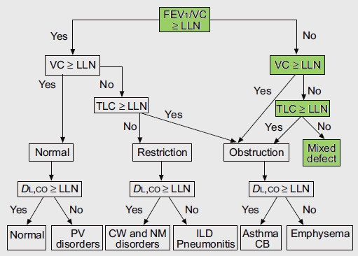
When I review spirometry results and either lung volumes or a DLCO test has also been performed, I will always check the Slow Vital Capacity(SVC) from the lung volumes and the Inspiratory Volume (Vinsp) from the DLCO test to see if they are larger than the reported Forced Vital Capacity. If either of them is I will manually re-calculate the FEV1/VC ratio to see if it indicates the presence of airway obstruction. This is in line with the ATS-ERS recommendations to use the largest Vital Capacity, regardless of the source, for the FEV1/VC ratio.

I have been reviewing the raw test data for all DLCO tests (as well as all the lung volume tests and regular spot checks on spirometry) performed in my lab for at least the last year. Since our software and hardware upgrade a year and a half ago we’ve found a number of problems that have significant effects on the DLCO test results. Depending on the problem they are capable of causing the results to be over- or under-estimated. All of the technicians performing the tests are now well aware of these problems and there haven’t been any problematic DLCO tests selected for a while. Nevertheless, I always check the raw data just to be sure.

Today, I ran across a report that looked quite straightforward. A set of spirometry and DLCO tests had been performed on a frequent-flier patient with pulmonary fibrosis. The patient has restrictive lung disease and lung volumes measured about a year ago were 64% of predicted. Even though the patient’s FVC has decreased since then there is no clinical reason to repeat the lung volume measurements. The results looked like this: 

  Observed: %Predicted: Predicted:
FVC (L): 2.28 51% 4.45
FEV1 (L): 1.64 51% 3.21
FEV1/FVC (%): 72 100% 72
DLCO ml/min/mmHg: 12.03 50% 24.23
Vinsp (L): 2.29    

Continue reading

The FEV3/FVC ratio, a useful tool for assessing early and mild airway obstruction

The FEV1 and FEV1/FVC ratio seems to have become the predominant, if not the sole factor for determining the presence of airway obstruction. It is true that a reduced FEV1/FVC ratio provides a strong and reliable signal for this purpose but its limitations have also been recognized for quite a while. The most obvious one is that the FEV1/FVC ratio will be falsely elevated when the FVC is underestimated. This is the primary factor driving the interest in FEV6 and the FEV1/FEV6 ratio. Less well appreciated is the fact that there are many causes and sites within the airways that can be involved in airway obstruction and that the focus on the FEV1/FVC ratio may cause certain forms of airway obstruction to be overlooked.

The FEF25-75 (aka MMEF) was originally proposed as way to determine the presence of small airways disease but it has since been shown to be an unreliable indicator. Most of the pulmonary physicians I work with have expressed doubt that there is such a thing as small airways disease but that doesn’t mean that some patients don’t have mild airway obstruction that is not evident when assessed solely by the FEV1/FVC ratio.

Continue reading

SVC and the FEV1/FVC Ratio

The Slow Vital Capacity (SVC) maneuver is usually performed as part of lung volume measurements. It is not unusual for the SVC to be larger than the FVC, particularly in patients with airway obstruction. This can have a bearing on the FEV1/FVC ratio and in fact the ATS-ERS recommendations for PFT interpretation say that the largest vital capacity value regardless of which test it comes from should be used to calculate the FEV1/VC ratio. When I review a full panel of tests (FVC, lung volumes, DLCO) I always check to see if the SVC or IVC (from the DLCO test) are larger than the FVC and then re-calculate the FEV1/FVC ratio and its percent predicted if they are. Test results that at first glance look normal will instead show airway obstruction often enough when this has been done that the time spent going through this process is worthwhile.

This only works however, when I have a full panel of tests to extract other vital capacities from. Patients that show airway obstruction when their FEV1/VC ratio is re-calculated have often had only spirometry performed on prior visits and their spirometry results were considered to be within normal limits at those times. Our lab software lets us select and report the “best” FVC and FEV1 from a series of spirometry efforts so this raises an interesting question and that is when and how often should a SVC maneuver be performed instead of a FVC maneuver during a spirometry session in order to get and report the largest VC?

Continue reading

Height and the GLFI FVC, FEV1 and FEV1/FVC ratio

Although the authors of the Global Lung Function Initiative (GLFI) study acknowledge the effect of height on their reference equations the range and distribution of heights in its study populations was not included in the report. This was a similar problem for the NHANESIII reference equations since the height range was never reported within the text of the original report however it did include scatter graphs showing the range of heights. These graphs imply the height range was 162 to 194 cm (64” to 76”) for caucasian males and 145 to 180 cm (57” to 71”) for caucasian females. Using the extremes of these height ranges it is interesting to see how the GLFI reference equations compare to the NHANESIII reference equations.

Continue reading

GLFI and the FVC, FEV1 and FEV1/FVC Ratio

The Global Lung Function Initiative (GLFI) was established by the European Respiratory Society in 2008 with the goal of establishing a truly worldwide set of reference equations for spirometry. Its results were released in the December 2012 issue of the European Respiratory Journal. Although the reference equations presently apply only to Caucasians, African-Americans and northern and southern Asians, it will likely be updated with Hispanic, African and Polynesian data within the next couple of years.

This has been a massive undertaking involving spirometry data from 72 different testing centers in 33 countries. The data has been subjected to rigorous quality control and an extensive, sophisticated statistical analysis and will likely become the standard reference equation set for spirometry testing in Pulmonary Function labs around the world.

Continue reading

The origin of FEV1 and the FEV1/FVC ratio

Over forty years ago when I started to learn about pulmonary function testing I was taught about the nuts and bolts of the tests, not where the tests came from or how they came into existence. Spirometry and the FVC, FEV1 and FEV1/FVC ratio have always appeared to be the core elements of pulmonary function testing and seemed most likely to have been scratched on the wall of the first paleolithic PFT lab cave. I have been reviewing a lot of older research papers lately, including several historical reviews, and was quite surprised to find out how recent both FEV1 and the FEV1/FVC ratio really are.

The first version of the modern spirometer (a counter-weighted volume displacement water seal spirometer) was developed by John Hutchinson in England around 1844. He performed vital capacity measurements on over 3000 people and in 1846 published a paper where he showed (among other things) the linear relationship of vital capacity to height. Different versions of Hutchinson’s spirometer were developed by other researchers in the following decades but even through the 1920’s the only measurement that was ever made with a spirometer remained just the vital capacity.

Continue reading