Anatomic dead space

I’d spent some time researching single-breath tests a while back and of course ran across the Fowler method for measuring anatomic dead space. It’s a relatively simple test but assessing its results as well as the results of alternate dead space measurement techniques turns out to be more complicated than I had remembered.

The official definition of anatomic dead space is that it is that part of the inhaled volume that remains in the airways at the end of inhalation and does not participate in gas exchange. An accurate estimate of this volume is important because respiratory dead space (Vd/Vt, discussed previously) is composed of both anatomical and physiological dead space. The physiological component of the respiratory dead space cannot be determined without knowing the anatomical dead space.

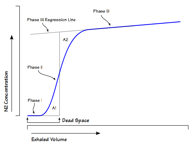
Anatomic dead space is usually considered to be the physical volume of the airways but static measurements of airway volume do not take into consideration the dynamic aspects of respiration. The most commonly used method for measuring anatomic dead space in a research setting is the single-breath technique developed by Fowler in 1948. In this method, after an inhalation of oxygen, the nitrogen concentration in an individual’s exhalation is plotted against exhaled volume.

Fowler Dead Space

Continue reading

Closing Volume

Closing Volume (CV) is a measurement made from a single-breath nitrogen washout (SBNW) test. It was commonly performed decades ago and elevated values were considered to be an indication of small airways disease and an aid in the detection of the early stages of airways disease. It is hardly ever performed any more but I still occasionally see research studies that include this test and almost every test system that is capable of measuring lung volumes by nitrogen washout is also capable of performing a CV.

The CV test is performed with a test system with an analyzer tap immediately next to the mouthpiece and a way of delivering 100% oxygen either from a demand valve or a reservoir. Originally this test was performed using a real-time nitrogen analyzer but it is now almost always performed with an oxygen analyzer instead. A subject is placed on the mouthpiece and exhales to RV and then inhales 100% oxygen to TLC. The subject then exhales steadily to RV and during the exhalation the subject’s exhaled nitrogen (either real or calculated from the oxygen concentration) is plotted against their exhaled volume and produces a curve that looks like this:

Closing Volume Graph

The trace is divided into four portions. Phase I is the very beginning of exhalation where only oxygen is being exhaled and consists primarily of test system and airway deadspace. Phase II is where the nitrogen concentration rises rapidly and consists of mixture of airway and alveolar gas. Phase III is where the nitrogen concentration plateaus and its slope depends on the uniform distribution of gas in the lung. Phase IV is where the nitrogen concentration rises more or less abruptly from the plateau and is considered to be part of the closing volume. The inflection point between phase III and phase IV is not always easy to discern and may need to be extrapolated from the phase III and phase IV slopes.

Continue reading