Diagnosing Mitochondrial Myopathies

I’ve been reviewing my CPET textbooks trying to get a better idea of how to differentiate between different cardiovascular limitations. The other day I ran across an article on a related subject and thought it might be instructive.

The hallmark of cardiovascular limitations is the inability to deliver enough oxygenated blood to the exercising muscles. Another limitation that has similarities to this (and one that is infrequently diagnosed) is the inability of the exercising muscle to utilize the oxygen delivered to it. The best examples of this type of exercise limitation are mitochondrial myopathies (MM).

The mitochondria are the primary source of the ATP used by exercising muscle. There are several conditions that can cause the number of mitochondria to be reduced and there is wide variety of mitochondrial genetic defects. Mitochondria have their own genes and these can have both inherited or acquired genetic defects which can cause anything from mild to severe decreases in the ability to produce ATP. Mitochondria require oxygen to produce ATP so when the number of mitochondria are reduced or their ability to produce ATP is reduced the rate at which oxygen is consumed by an exercising muscle is also reduced.

A relatively common complaint of individuals with MM is dyspnea and exercise intolerance. One study found that 8.5% of all the patients referred to a dyspnea clinic had a mitochondrial myopathy of one kind or another. A definitive diagnosis requires a muscle biopsy but because the symptoms are often non-specific and a biopsy is an invasive procedure, it is usually not performed unless there more significant evidence suggesting a MM.

Continue reading

What does it mean when Ve exceeds its predicted during a CPET?

When I review the results from a CPET I am used to considering a maximum minute ventilation (Ve) greater than 85% of predicted as an indication of a pulmonary mechanical limitation. Recently a CPET report came across my desk with a maximum minute ventilation that was 142% of predicted. How is this possible and does it indicate a pulmonary mechanical limitation or not?

It is unusual to see a Ve that is greater than 100% of predicted. We derive our predicted max Ve from baseline spirometry and calculate it using FEV1 x 40. We have tried performing pre-exercise MVV tests in the past and using the maximum observed MVV as the predicted maximum Ve but our experience with this has been poor. Patients often have difficulty performing the MVV test correctly and realistically even if it is performed well the breathing maneuver used during an MVV test is not the same as what occurs during exercise. Since both Wasserman and the ATS/ACCP statement on cardiopulmonary exercise testing recommend the use of FEV1 x 35 or FEV1 x 40 as the predicted maximum minute ventilation we no longer use the MVV.

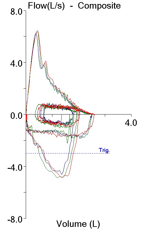
There are usually only two situations where a patient’s exercise Ve is greater than their predicted max Ve. First, when a patient is severely obstructed their FEV1 is quite low and FEV1 x 40 may underestimate what they are capable of since they are occasionally able to reach a Ve a couple of liters per minute higher than we expected. Second, if the FEV1 is underestimated due to poor test quality then the predicted max Ve will also be underestimated. In this case however, the baseline spirometry had good quality, was repeatable and the results did not show severe obstruction but instead looked more like mild restriction.

Pre_Exercise_FVLs

Effort 1: Effort 2: Effort 3:
FVC (L): 2.51 2.52 2.60
FEV1 (L): 1.86 1.87 1.95
FEV1/FVC %: 74 74 75
PEF: 6.26 6.46 6.37

Continue reading

Assessing MVV results

The Maximum Voluntary Ventilation (MVV) test was initially described in 1933. It was the first pulmonary function test that involved inspiratory and expiratory air flow in a significant way and for this reason it helped to set the stage both conceptually and technically for the FEV1, the FEV1/FVC ratio and our present understanding of obstructive lung diseases. MVV is reduced in a variety of conditions, including obstructive, restrictive and neuromuscular diseases, but a reduced MVV is non-specific and this limits its clinical utility. Nevertheless, it continues to be performed both in clinical labs and for research, and for this reason it would seem to be a good idea to know how to assess MVV results.

As usual, there are two aspects to assessing pulmonary function results; test performance and normal values.

Currently the ATS/ERS statement on spirometry contains the only available standard for performing the MVV test. Unfortunately this standard also contains some significant flaws. Its primary recommendation is that the MVV test be performed with a tidal volume that is approximately 50% of the forced vital capacity and a breathing frequency of around 90 breaths per minute. These recommended values are problematic and some simple mathematics will show why.

A respiratory rate of 90 BPM means that there is 2/3 of a second for both inhalation and exhalation. With a 1:1 ratio for inspiration and expiration, there is only 1/3 of a second for exhalation. Since it normally takes a full second to exhale approximately 75% of the vital capacity (i.e. the FEV1), 1/3 of a second would only allow time to exhale 25% of the vital capacity (not exactly true of course, but it helps prove the point). How then is it possible to exhale 50% of the vital capacity, twice that amount, in the same amount of time? The answer is that it isn’t and if it was somehow possible for somebody to actually meet the ATS/ERS recommended values they would have an MVV that would be 45% to 100% higher than any of the predicted MVV’s. I suspect the ATS/ERS agrees this is a problem since following the initial recommendation it also says that “… since there are little data on MVV acceptability criteria, no specific breathing frequency or volume is required”.

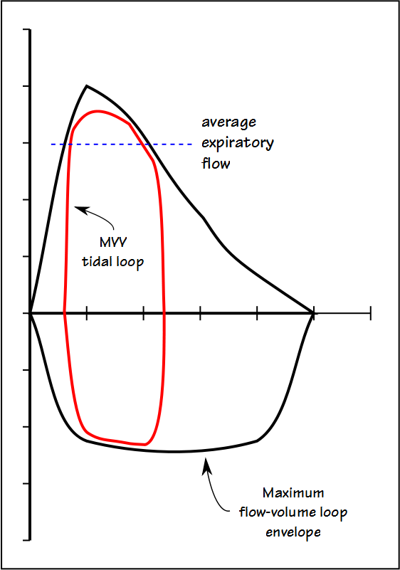
The fact is that no single tidal volume recommendation is going to work for all patients and this is because the MVV tidal volume has to reside mostly within the maximal flow-volume loop envelope.

MVV_TV_FVL_01

Continue reading

Is the MVV clinically relevant?

The respiratory system is in part a mechanical pump or bellows. The Maximum Voluntary Ventilation test (MVV, aka Maximum Breathing Capacity, MBC) is intended to measure the maximum ventilation a patient is capable of. As such the results are dependent on a patient’s lung volume, respiratory muscle strength and endurance, airway resistance and overall inertia of the thoracic cage.

When I started doing PFT’s in the early 1970’s the MVV was a standard part of a complete workup. This has long since changed and I have not performed the MVV test routinely in over 25 years but I’ve always wondered what the MVV test is actually supposed be measuring in a clinical sense.

The ATS/ERS statement on spirometry recommends that the MVV test be 12 seconds long and that for optimum results the patient’s tidal volume should be approximately 50% of their VC at a respiratory rate of 90 breaths per minute. Tidal volume is accumulated during exhalation and at the end of the test the accumulated volume is then multiplied by 5. The ATS/ERS statement also suggests that MVV values that are less than FEV1 (L) x 40 indicate suboptimal patient effort.

Continue reading