Post-BD FVC. It’s about time.

When assessing the response to bronchodilators the change in FEV1 is used far more frequently than any other spirometry result. Other values such as inspiratory capacity (IC) and peak inspiratory flow (PIF) have also been proposed as indicators, but the ATS/ERS standards includes changes in FVC as well as changes in FEV1 and this is often overlooked. Specifically they:

…recommend using the per cent change from baseline and absolute changes in FEV1 and/or FVC in an individual subject to identify a positive bronchodilator response. Values >12% and 200 mL compared with baseline during a single testing session suggest a ‘‘significant’’ bronchodilatation.”

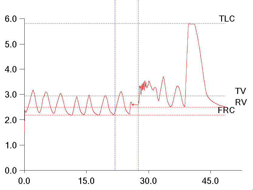
I don’t have any particular disagreement with this since post-BD increases in FVC are probably similar in nature to the post-BD changes in IC seen in some individuals with COPD. So when spirometry results like this:
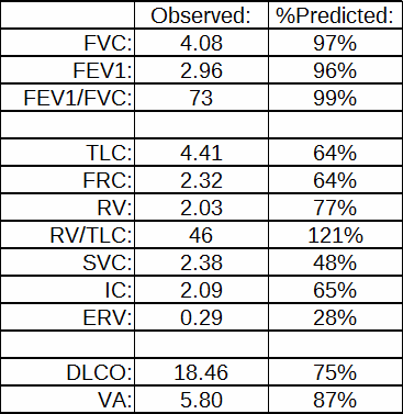
Pre-BD: %Predicted: Post-BD: %Change:
FVC: 1.82 66% 2.55 +40%
FEV1: 0.66 32% 0.72 +10%
FEV1/FVC: 37 49% 29 -22%

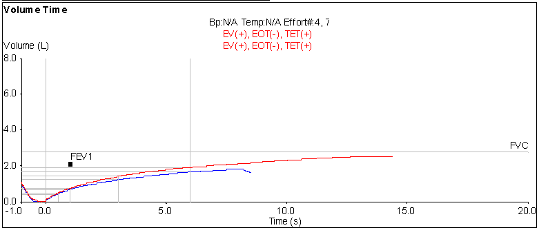
comes across my desk, I’m inclined to consider that the results show a positive bronchodilator response. Post-BD increases in FVC are not usually quite as large as 40% however, so I took a closer look at this particular test. When I did what I saw was that the post-BD test length was significantly longer than the pre-BD test length.

Longer_FVC

Continue reading

Helium overshoot, revisited

A while back one of our technicians brought a helium dilution FRC graph to my attention and wanted to know if it showed a system leak. At that time my response was that it definitely wasn’t a leak (leaks don’t show increases in helium) and was probably due to too much oxygen being added to the system at the beginning of the test.

Helium_Overshoot_01

A couple of days ago a technician brought a similar graph to me and again I was asked why it looked unusual. I’ve had time to think about this issue since the last time and I’ve come up with an alternate explanation that I think fits the facts a bit better.

A normal helium dilution curve looks something like this:

Helium_Overshoot_02_nl-ish

which shows the helium decreasing with what is more or less an exponential decay curve. What’s unusual about the other curve is that it shows a relatively rapid fall to the lowest helium concentration near the beginning of the test and then a slow rise to the final concentration.

Continue reading

Adjusting DLCO for hemoglobin

My hospital’s Oncology division treats a number of patients with lymphoma and leukemia. It also has an active bone-marrow transplant program and for all of these patients diffusing capacity measurements are a critical part of assessing treatment progress. Since these patients are also frequently anemic, correcting DLCO results for hemoglobin is also critical.

For a factor that has as much importance for the interpretation of DLCO results as it does the effect of hemoglobin on DLCO has actually been studied a relatively small number of times. Part of the reason for this is the problem of finding an acceptable model. A reduced or elevated hemoglobin is a consequence of many diseases and conditions. When studying patients longitudinally it is often difficult to separate the changes in DLCO that occur from the disease process and those that occur from changes in hemoglobin. For this reason changes in hemoglobin pre- and post-treatment in anemia and polycythemia have been studied most frequently.

The ATS/ERS currently recommends correcting DLCO for hemoglobin (although notably they recommend that the predicted DLCO be corrected, not the observed value) using the equations developed by Cotes et al in 1972. Cotes’ work was based on subjects with iron-defficienty anemia but just as importantly on theoretical considerations involving Roughton and Forster’s equation on the relationship between the membrane and hemoglobin components of the diffusing capacity:

1_over_DLCO_formula

Cotes

Continue reading

A real fixer-upper

I was reviewing reports today when I ran across one with some glaring errors. There were several things that immediately told me that the reported plethysmographic lung volumes were way off; the VA from the DLCO was almost a liter and a half larger than the TLC and the SVC was only about half the volume of the FVC.

Table1

When I took a look at the raw test data I saw at least part of the reason why the technician had selected these results to be reported and that was because the SVC quality from most of the efforts was poor. They mostly looked like this:

Fixer_Upper_01

It is apparent that the patient leaked while panting against the closed shutter and this caused the FRC baseline to shift upwards. I’ve discussed this problem previously, but when this happens the RV is larger than the FRC, there is a negative ERV and the TLC is overestimated. There is no way to fix this problem from within the software. The FRC is determined by the tidal breathing before the shutter closes and cannot be re-measured afterward.

Continue reading