Anatomic dead space

I’d spent some time researching single-breath tests a while back and of course ran across the Fowler method for measuring anatomic dead space. It’s a relatively simple test but assessing its results as well as the results of alternate dead space measurement techniques turns out to be more complicated than I had remembered.

The official definition of anatomic dead space is that it is that part of the inhaled volume that remains in the airways at the end of inhalation and does not participate in gas exchange. An accurate estimate of this volume is important because respiratory dead space (Vd/Vt, discussed previously) is composed of both anatomical and physiological dead space. The physiological component of the respiratory dead space cannot be determined without knowing the anatomical dead space.

Anatomic dead space is usually considered to be the physical volume of the airways but static measurements of airway volume do not take into consideration the dynamic aspects of respiration. The most commonly used method for measuring anatomic dead space in a research setting is the single-breath technique developed by Fowler in 1948. In this method, after an inhalation of oxygen, the nitrogen concentration in an individual’s exhalation is plotted against exhaled volume.

Fowler Dead Space

Continue reading

Proposal to improve the readability of flow-volume loops

I’ve been planning on putting together a tutorial on characterizing and interpreting the contours of flow-volume loops so I’ve been accumulating flow-volume loops that are examples of different conditions. Lately I was reviewing some of them and noticed that when I tried to compare loops from different individuals with similar baseline conditions that the different sizes of the flow-volume made this difficult. For example, these two loops are both from individuals with normal spirometry.

FVL_Scaling_05

FVL_Scaling_08

One is from short, elderly female and one is from a tall, young male. If all you had to look at was the flow-volume loops, you might think that the smaller loop was abnormal, but the larger loop actually comes from a spirometry effort with an FVC that was 92% of predicted while the smaller loop’s FVC was 113% of predicted. The difference in sizes of these loops is of course due to the difference in age, gender and height between these individuals but also because of settings we’ve made in our lab software and because of the ATS/ERS spirometry standards.

Continue reading

DLNO isn’t the same as DMCO but sometimes it’s useful to pretend it (almost) is

Oxygen transport between the lungs and the body depends on numerous complex factors. Ventilation and the alveolar-capillary surface area are of course important but a critical component is hemoglobin. Oxygen is poorly soluble in water (which is what blood is mostly made of) and the transportation of oxygen throughout the body would not happen without hemoglobin’s ability to absorb and release oxygen on demand. Although it is possible to measure the diffusing capacity of oxygen (DLO2) the process is technically difficult and not at all suited to routine clinical testing.

There are a number of gases that are able to diffuse across the alveolar-capillary membrane and can be used in a variety of physiological measurements but in order for a particular gas to act as a substitute for oxygen it must be able to interact with hemoglobin. Carbon monoxide (CO) has an affinity for hemoglobin approximately 220 times greater than oxygen and was the first gas used to measure diffusing capacity (DLCO). DLCO has been a routine test for well over 50 years and has been measured by single-breath, steady-state and rebreathing techniques.

Nitric Oxide (NO) has an affinity for hemoglobin about 400 times greater than carbon monoxide (it is generally an irreversible process since the end product is methemoglobin whereas hemoglobin’s binding with CO is more reversible) and for this reason it can also be used to measure diffusing capacity. DLNO can also be measured by single-breath, steady-state and rebreathing techniques. Because of its high affinity and the speed at which the binding of NO to hemoglobin occurs numerous researchers have assumed that DLNO is equivalent to DMNO (the membrane component of diffusing capacity). This is not really true, but it can be a useful fiction and in order to understand why it’s necessary to look at the basic physiology of diffusing capacity tests.

Roughton and Forster’s seminal 1957 paper showed that diffusion is the sum of two resistances. I’ve discussed this previously but specifically:

1_over_DLCO_formula

Where:

DMCO = membrane component

θCO = the rate at which CO binds to hemoglobin

Vc = pulmonary capillary blood volume

The first resistance (1/DMCO) is the resistance to the diffusion of CO through the alveolar-capillary membrane and blood plasma to the surface of the stagnant plasma boundary layer around a red blood cell. The second resistance refers to the diffusion rate of CO through the plasma boundary layer, then the wall and interior of the red blood cell and finally the rate of reaction with hemoglobin.

DL_Resistances Continue reading

Alveolar O2 and Altitude

Recently I was trying to explain the effect of altitude on blood oxygenation to somebody with IPF. They had observed that their oxygen saturation was fairly normal at sea level but that they needed to use their supplemental O2 when they went up to 2000 feet and didn’t understand why such a low altitude made that much of a difference. I don’t think I did very well with the explanation since at the time I was limited to only text and I much prefer pulling out diagrams and waving my hands in the air.

The place to start is with the alveolar air equation.

Alveolar Air Equation 1

Where:

PAO2 = partial pressure of O2 in the alveoli

FiO2 = fractional concentration of O2

PB = barometric pressure

PH20 = partial pressure of water vapor in the alveoli

PaCO2 = partial pressure of CO2 in the arteries

RER = respiratory exchange ratio (VCO2/VO2)

When normal values are plugged into the alveolar air equation it looks like this:

Alveolar Air Equation 2

An important point is that when air is inhaled into the lung oxygen is diluted by the water vapor and carbon dioxide that are already there. The partial pressure of oxygen will vary with atmospheric pressure but the partial pressure of water vapor and carbon dioxide are relatively fixed values. Atmospheric pressure of course decreases with altitude.

BP_Altitude

Continue reading

What’s in a name?

My lab is in the final stages of a software update that will allow for electronic signing of our reports. This has been a long and slow process partly because the release date of the software got pushed back several times but mostly because a wide variety of different hospital departments and sub-departments have had to be involved.

In all the years that I’ve had computers in the pulmonary function lab I’ve never gone through a software update that was either as easy as expected or occurred within the original schedule. This includes the time when all we had was a single IBM PC/AT with a 40 megabyte hard drive, no network and the only people that cared we were going through an update was ourselves. Since we now have a dozen networked PCs located in two different building on-campus as well as three off-site locations using an IS-managed SQL server and HL7 interface I didn’t have any expectations for a speedy update and so far I have not been disappointed.

This time because the update revolves around electronic signing the hospital’s Health Information Management (HIM, i.e. Medical Records) department has been significantly involved. Among other things this has led to HIM reviewing all of our reports and requiring changes to bring them up to hospital standards. To some extent this make sense since, for example, they require that patient identification be exactly the same on all reports from all departments (same fields, same locations).

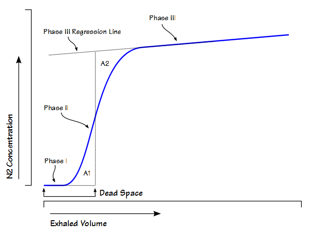
However, they also questioned some of the terminology used on our test reports. We’ve used the default test names that were in our report format editor (yes, we’re that lazy) and until they were brought to our attention I never really thought how odd some of them were. In particular, some of the terms used for the diffusing capacity didn’t make a lot of sense. For example, DLCO corrected for hemoglobin was DsbHb and DLCO/VA was reported as D/Vasbhb. To some extent I understand where these names came from but the reality is that they are in part holdovers from the past, in part they come from a need to keep names short so they fit in what space is usually available on reports, and in some cases they were probably created by programmers who hadn’t the slightest idea what the correct nomenclature should have been.

Note: Dsb likely comes from a time when you needed to differentiate between the results of different types of DLCO tests (steady-state and single-breath). Since there hasn’t been a test system built for at least 40 years that could perform a steady-state DLCO, the need to make this distinction is long since past.

Continue reading